 Surgically
enhanced venomous snakes. Venom glands out, silicone implants in!� The creation of perfect exhibition snakes in
the post HIH era.
Surgically
enhanced venomous snakes. Venom glands out, silicone implants in!� The creation of perfect exhibition snakes in
the post HIH era.
Raymond Hoser
488 Park Road, Park Orchards,Victoria, 3114, Australia.
Email:
adder@smuggled.com
Originally Published in CROCODILIAN:JOURNAL OF THE VAAH
5(1)(May 2005):17-28,5(2)(August 2005):17-28 (and covers),5(3)(November 2005):30-36.
Abstract
This paper follows
on from the report of the first successful series of operations on Australian
elapid snakes to make them venomoid as published by Hoser (2004) and also
reprinted in Crocodilian in 2004.
It recaps the
original series of operations as well as the first successful operations in
Australia using silicone implants as a means to make the operation effectively
undetectable in larger snakes.
All operations
conducted were 100% successful in that the snakes made full recovery within
incredibly short time frames and in that the final result was effectively
undetectable to casual observers.� More
than a year after these operations, all snakes remain alive and in the peak of
physical health.
Silicone implants
are effective at masking the finished procedure in large mature snakes with
sizeable venom glands and where removal of them may result in a noticeable
thinning of the head.
The procedures
given here should be used as a model for others intending performing such
surgery on dangerously venomous snakes.
Published
criticisms of the original Hoser operations, as made by habitual critics of
anything Raymond Hoser does, are addressed and found to lack merit as are the
popularly touted arguments against venomoid surgery per se.
In future the
driver of venomoid surgery will be for the benefit of snakes, not their
keepers.
INTRODUCTION (WHY
BOTHER?)
In my own case, I
have been keeping and handling deadly Australian snakes for some decades and
never had a call to remove the venom glands from snakes.
The initial push to
remove venom glands came from several angles and arose due to my seeking a
permit to publicly display, show and handle deadly species of snake and
minimize the inherent risks, both real and perceived.
Noting the increase
in public liability insurance problems and related occupational health and
safety laws, particularly in the wake of insurance disasters in Australia such
as the Longford Gas Explosion, HIH Insurance Company bust up and so on the idea
of using deadly snakes for displays in close quarters became problematic.
In the period
2002-2004 I received inquiries from numerous people who wanted a live and
interactive display of deadly snakes, but without the associated risks.� Included here were the managers of the
Heritage Golf Club at Chirnside Park (Victoria), Diamond Creek Public School at
Diamond Creek, the Bayside Christian College at Baxter and others.
In other words
these people wanted to have their cake and eat it too.
Whilst in the past
it was easy to tell these people to simply accept the possibility of a
snakebite risk, recent events here in Australia were changing things.
In the 1990's a
respected (but then young) keeper Aaron Briggs from Coburg, (Melbourne,
Australia) was bitten by a pet Death Adder (Acanthophis antarcticus).� That in itself wasn't too exceptional, but
for the fact that it was a quiet news day and the local news media got onto the
story.
Within days the
"do gooders" were demanding that reptile keeping be outlawed and the
herpetological community had to fight a rearguard action to preserve our hard
won rights.
Ultimately, the
keeping of venomous snakes was made more restrictive (including outlawed to
under 18's), but no other rights were lost.
Just prior to doing
a snake show in 2001, Fred Rossignolli was bitten by a large King Brown Snake (Cannia
australis).� Instead of doing a
snake show, he landed at the Maroondah District Hospital and again there were
calls to outlaw the keeping and displaying of venomous snakes.� Similar happened after he got a bite from a
Copperhead during a show at a teacher's conference and he had to cut things
short.
The calls for
further restrictions blew over and nothing significant changed, but it was a
wake-up call to those who keep or handle deadly snakes.
Regulation of
exhibitors did however tighten up to include the need for more secure
(lockable) cages/containers, warning labels and the like.
In the 2002
Melbourne Agricultural Show, Workcover officials whinged at Fred Rossignolli
for walking barefoot in his pit full of dangerously venomous snakes.�
Fred wasn't allowed
to show snakes at the 2003 show, even though his show was generally regarded as
"the best" and no incidents occurred the previous year (bearing in
mind Fred does such shows daily and has only been bitten once in ten years
doing such shows).
Yes, he's had a few
"food bites" at home, but that's another story.
Another snake
handler, Simon Watharow, "stole" the Melbourne show gig from
Rossignolli on the basis that his display was "safer", even though it
was generally conceded that the entertainment value and educational benefit of
the Watharow show was vastly inferior to Rossignolli's.
(Rossignolli free
handles his snakes, while Watharow uses pinning sticks, cripple tongs and hooks
only, the result being an inferior view of the snakes by the audience).
The following year,
Watharow managed to again get the Melbourne show gig by defaming rivals with
false statements, even after one had been booked, resulting in a cancellation.
In other words,
there was a push from persons and organisations outside of myself for a safe
alternative to educating the public with deadly snakes, with all the benefits
of the handler being able to free handle snakes as needed, but at the same time
to minimize the risks.
Internet forums are
rife with comments about venomoid snakes (generally negative) and I am not
going to add my own views to the already polarised views online already, save
to dispel a few myths under the next heading and elsewhere in this paper.
However, having
been involved in snake science for more than 3 decades, I can assure readers
that my own handling skills are sufficient to exclude the need for me to neuter
the venom glands of snakes for my own ends.
MYTHS AND
CORRECTIONS
Venomoid is the
term used for snakes who have had their venom glands removed and the name given
to surgical procedures to do this.
From a practical
point of view, snake venom is of no discernable benefit in terms of digesting
food.
This is evidenced
by the fact that pre-killed food is digested just as easily (and rapidly) as
food killed by snake venom.
In other words any
breakdown of tissues of prey by venom is of insignificant benefit in terms of
aiding digestion, or being essential to it.
Claims to the
contrary have no substantive basis.
If contrary claims
did have a factual basis, then it would not be possible to feed venomous snakes
indefinitely on pre-killed food, or in the case of some Australian elapids,
such as Tiger Snakes (Notechis scutatus) a diet of items as diverse as
dead fish, steak, sausages, chicken slabs or chicken necks, pork, ham and
Calamari (squid), all of which I have done.
Similar applies for
other elapids, fed nearly as varied diets, including Red-bellied Black Snakes (Pseudechis
porphyriacus), eastern Brown Snakes (Pseudonaja textilis),
Small-eyed Snake (Rhinoplocephalus nigrescens), Death Adders (Acanthophis
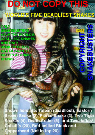
spp.), Collett's Snakes (Panacedechis colletti), Taipans (Oxyuranus
scutellatus) and others.
Removal of venom
glands in other words is merely the neutering of the snake's ability to kill
live prey.� As the process is not
reversible this means that the snake can (probably) not be fed live food again.
In the captive
situation of keepers like myself, this is of no relevance as food given to all
snakes is dead and taken from a freezer.�
It does however mean that the snake cannot be released into the wild and
expected to fend for itself.
But the inability
to immobilize live prey is the only measurable negative of venomoid surgery and
in the real world of herp keepers is rarely an issue.
Performed
correctly, venomoid surgery is not particularly painful for the snake and
recovery from the surgery is very rapid.�
This is amply demonstrated later in this paper.
As only
non-essential soft tissue is excised, recovery speed is fast due to the fact
that no bone or tooth repair is (usually) necessary and healing tissue is
generally fixed (not moving) and not in use as would be the case for something
like a limb in a human.
Snakes on which
surgery is performed (properly) will often be willing and able to take food
almost immediately after operation, as in taking food offered within days.
Complications from
surgery in the form of injury, infection or arising from anesthesia in properly
performed operations are almost unknown (and totally unknown in my own
situation) and in the rare cases where they may arise, are (presumably) easily
treated and dealt with.
Venomoid surgery is
not essential to snakes in that it is not necessary to save the snake's life.
It is best classed
as "elective surgery" and hence should only be undertaken on healthy
well-adjusted snakes in circumstances where there is no known risk to the snake
from such a procedure.
It should be
treated in much the same light as (sexually) neutering a dog or cat, although
the latter operations have far greater long-term effects on the health and
personality of the subject animals.
WHO SHOULD PERFORM
VENOMOID SURGERY ON SNAKES
As a procedure it
is remarkably simple and while it would be generally advised that a qualified
veterinary surgeon perform the operation, this is not necessarily essential or
for that matter the most important requirement.
What is more
important (and far more so) is that the operation is only performed by a person
experienced in performing such operations and who is familiar with the exact
procedure to be followed in terms of what must be done.
This experience can
only be gained by doing such operations and should therefore be gained in the
first instance by studying of appropriate dead snakes upon which the operation
can be practiced as often as is necessary.
Sedation procedures
used to neutralise the snake during the operation should be fully tested on
appropriate snakes prior to doing a "real" operation, so that nothing
is left to chance when the first operation on a live snake is performed.
The operation
should only be performed by a person familiar with the snake species to be
operated on and who is skilled at handling the reptiles.
Noting the
pre-operational matters of sedation and after the operation itself, revival, it
is essential that the operation be performed by a person who is skilled and
experienced at handling the said species.
This paper explains
a successful process used and refined, so that if and when others need to
perform such surgery, that it can be done using proven and tested methods that
will not adversely affect the snake/s in question.
VENOMOID SURGERY
The species in
question here to be 'neutered' so to speak, were (in the first instance) Tiger
(Notechis scutatus), Eastern Brown (Pseudonaja textilis) and
Copperhead (Austrelaps superbus).�
All are deadly venomous snakes and the first two account for the vast
majority of fatal bites in Australia, including among keepers and non-keepers.
The only record of
venomoid surgery in Australia was the case published by Dave Millar in Herpetofauna
in 1976.
In that case
involving two Tiger Snakes, he made an incision into the side of the head (from
outside the head) and removed the gland immediately underneath.
Millar used various
methods to immobilize the snakes during the procedure and for reasons unknown
did two operations on each of two Tiger Snakes, removing one gland at a time.
For the first
operations, the snakes were immobilized by cooling to a state or torpor and
then by being held down at the relevant temperature were operated on.
The snakes
apparently healed well and both accepted food a week later.
In the second
operations, to remove the second venom glands the snakes were immobilized with chloroform.
This time the operations were not a success.� One animal developed a series of (linked?) infections and died,
while the other had a somewhat checkered road to recovery.
It was presumed that the problems arose from the sedation process and not
the surgery itself, but this isn't certain.
Either way, it is clear that the Millar operations were an abject
failure.
While there are frequent claims made about venomoid surgery by reptile
keepers, there has been no proper paper detailing a tried, tested and
apparently risk-free procedure for doing the operation and so before commencing
the first operation further research was required.
Consultation with veterinary surgeons and the relevant texts (such as Fry
1991 and Mader 1996), revealed no shortage of ways to sedate and/or anesthetize
snakes and immobilize them.
However a common complaint by various veterinary surgeons was the
differential between snakes in terms of required dosage needed to immobilize
the snakes, even of the same size and species.
Related to this was the fact that the margin for error in such procedures
was not always great and while with experience, losses of snakes under
anesthesia are not great (especially when using Isoflurane gas), there always
remained a risk.
A second issue was the problem of snakes reviving during an operation
after the sedation drug wore off.� No
drug treatment could remove the risk of this problem entirely.
To the contrary, cooling a snake above it's known thermal minimum didn't
seem to present any problems (see below for an exceptional case) and based on
the Millar case (above), it seemed to be the preferred means of immobilizing
snakes for the operation.
Several veterinary surgeons recommended the use of a modeling clay mould
with "arms" to hold down and immobilize sedated reptiles when surgery
was to be performed.
In the first instance, a variant of this was planned to perform this
operation, but it was ruled inferior to the final means (explained below), as
even with clay to restrain a snake, it was possible for a partly sedated snake
to be able to squirm it's way out of the holder.
Bearing this in mind, in preparing for the surgery in my case/s, a far
more effective means was devised to restrain snakes without incident.� This is explained later and shown in the
photographs taken at the time and in the absence of other yet unknown better
procedures, I strongly recommend that persons doing venomoid surgery use the
techniques developed and refined by myself as given in this paper.
The method I devised allowed me to immobilize any species of snake
without causing them harm, keep them immobilized as long as necessary and then
to allow them to revive almost immediately after surgery.
My means of sedation of snakes was via cooling to a temperature
sufficient to immobilize the snake, but not to be life threatening.
Tests on (Melbourne) Tiger Snakes, established that they could be cooled
to 5 degrees Celsius without incident. At this temperature they were
effectively immobile and could be manipulated almost as one pleased.
Fry 1991 (p. 421) mentioned that to maintain this temperature on reptiles
during surgery (4 degrees in his case), ice-baths could be used.
Claims that cooled snakes were abnormally prone to ailments if cooled in
this manner (see Fry 1991), did not correlate with my own experience in terms
of cooling reptiles in fridges for photographing.
Put simply, the said reptiles recovered perfectly and with no ill
effects, long or short term!
My own view is that authors citing cooling as cause of disease have
confused short term cooling with long term keeping at sub-optimal temperatures,
which is an issue unrelated to surgery or this paper.
Once a means had been developed to sedate snakes, the only remaining
hurdle was the means by which to conduct the surgery.
But before I explain this, I will outline some essential facts about the
snake's venom glands.
The venom glands are located above the jawline and generally posterior to
the eye to about the back of the skull, usually in the vicinity of and level
with the end of the mouth line, or slightly past this and slightly above.
This seems to be the positioning in all venomous snakes, including for
example the Pacific Rattlesnake (Crotalus viridis oreganus) as shown in
Fry 1991 (page 450, top three photos), indicating that venom glands and
delivery means (gland, duct, fang) in snakes only evolved once.
The glands (one on each side of the head) are sited under the rear head
shields and surrounded by muscle tissue, although in many elapids the amount of
muscle present between the glands and the scales is very little.� The glands generally sit outside the jawline
and with the surrounding muscle tissue form a major part of the flesh in the
posterior dorsal head region around the back of the skull.� In non-venomous snakes, such as pythons, the
equivalent flesh is either fat or muscle tissue, making them have heads of
roughly similar shape.
The exact size and positioning of the venom glands varies from snake to
snake, but appears to be larger (and reaching further back) in larger
non-growing snakes of a given species.
To the rear, the gland has a rounded end, joined to a stalk of muscle,
while anteriorly it narrows to form a vessel or duct that runs under the jaw
and into the fang.� The length of the
venom duct in terms of it's run from the venom gland to the fang tooth also varies
from snake to snake.
It is not always distinct, in that the narrowing may be fairly rapid, or
in some snakes more gradual and this variation appears to even occur within a
given species and even with age or size.
In some snakes the duct is up to 1 cm in length and very obvious as a
duct, while in some snakes it appears to be almost indistinct with the venom
gland almost appearing to narrow and run into the fang.
While more-or-less pointed at one end and with a blunt end at the
posterior end, the glands are more-or less rectangular (if viewed from above),
being cylindrical in shape, and encased with muscle tissue which in tandem with
the muscle stalk at the rear, appear to help push the venom to the fang.� (Expulsion of venom appears to be when the
muscles surrounding the sheath of the fang are depressed as happens when the
snake bites on something).
This muscle tissue is fairly easy to separate from the venom glands along
the length of the glands, but at the rear, both gland and muscle is affixed to
the rear of the head or the flesh of the neck.�
To separate this, one must cut it.
At the anterior end of the gland, the venom duct must also be cut to
remove the gland, and when doing so, it is important to cut the duct and not
the anterior part of the gland (otherwise leaving part in the snake).
While a number of texts detail the structure of venom glands in snakes,
preceding surgery it was essential for me to dissect snakes and look at these
structures for myself.
Initially it was envisaged that I'd dissect dead captive snakes as
"test runs" for surgery", but in late 2003, I was fortunate to
find several road-killed Tiger Snakes in order to inspect them.
While some corpses were quite decayed and smelly, they were still
adequate for me to inspect the venom glands and test means of conducting
surgery, instruments to be used and even refine the means by which I eventually
immobilized the snakes in surgery.
Tested were both the "external excision" method, by going
through the side of the head, and the "internal excision" method, of
going through the roof of the mouth, which was ultimately decided to be the
preferred method.
It was only after all aspects of surgery had been addressed and tested as
best as possible on dead snakes that an operation was conducted on a live
snake, which had already been immobilized and kept so for a period equal to or
longer than an operation would take and recovered without incident.
Before detailing the first venomoid operation, I should mention that I
subsequently developed an effectively foolproof method of putting reptiles (of
any species) into a state of cold torpor without causing risk to them and this
will be detailed later.
THE FIRST VENOMOID OPERATION
The method used for this operation was the same as that used for all
others, save for the manner of cooling and minor refinements to the means of
restraining the reptiles.
The venom glands were removed by an operation into the roof of the mouth
to remove them internally, the result being no cutting to the external surfaces
or scales of the head.
The subject was a half-grown Tiger Snake.� It was put in the fridge to get it's temperature to the desired 5
degrees Celsius, which was the setting in the fridge.
The snake was in a state of torpor, but obviously still alive.
It was then quickly removed from the fridge and placed upside down on a
60 cm long wooden plank (removed from the fridge) and then by it's head and
snout sticky taped down to the wood, with the snout and neck at a predetermined
spot and held down.
The tape ran over the head only.
The following section of the neck was quickly sticky-taped down to the
wood (with the tape running around the board in full circles), with the rest of
the snake then being unrestrained but placed over an adjacent towel.
The area near the heart (about 1/8 of the way down a snake's total
length) was not restrained in any way (nor was lower down the snake's body in
this operation, but for later operations lower down was restrained as well, as
sometimes this moved away from or out of contact of the towels and the further
restraint prevented this).
The towel (just mentioned) had previously been folded, wet with water and
frozen solid.� Before the operation it
had been allowed to thaw to the extent that it was still mainly frozen (in the
core) but the surface was wet and it could also be bent or moved with some
difficulty.
In other words it was pliable.
(At a room temp. of about 20�C it
takes about an hour for a towel to reach this state and it remains OK for about
an hour and by rotating towels, a supply can be maintained indefinitely).
A second similar towel was placed over the snake's body, forming a cold
blanket above and below the snake.
This design allowed me to maintain the cold torpor during the course of
the operation, but not actually freezing the snake.
The tape over the snake's head and snout was then removed (having been in
place for only some seconds), with the snake itself opening it's mouth to
breath.
(For those unaware, the glottis, or windpipe of a snake is not always
open.� It opens and shuts periodically
and a momentary blockage is not a fatal condition, so long as it is kept just
that, momentary).
The lower jaw and upper jaw were then fixed in a position to allow
surgery to start.
Veterinary surgeons suggested using string, sutures and other materials
to affix the snake's jaws during surgery, but after some previous testing on
the snake (without actually doing surgery), it'd been established that the best
means to affix the jaws and head in place was as follows.
The neck region had already been fixed flat and in a straight line with
sticky tape.
By it's nature, this effectively prevented the snake from any means to
squirm loose, even if it were to regain consciousness or an ability to move
during the operation, which may occur if the snake were insufficiently cooled.
No other effective way was found to properly restrain the snake.
To either side of the head and neck region of the snake and already
affixed to the wood plank, were nails.�
These had been affixed as contact points for so-called "twist
ties".
These are thin metal strips that can be easily bent and twisted to form a
tight line or knot.
A long strip was used to affix the lower jaw to the wood, while a second
strip was used to do the same to the upper jaw, making sure that the glottis
(windpipe) remained clear.
In a breathing snake, this periodically opens and shuts and this remains
the case in the snake as it is operated on.
A hard-wire frame was set between two nails to hold up the lower jaw (on
top) to keep the mouth open for the operation.�
The nails were set (slightly in facing) to make the frame naturally rise
and fix in position, thereby holding the upper jaw in place and fixed.
Nails on the board (several on each side) were spaced apart to allow for
any snake to the size of a three metre elapid and to allow fixing of more than
one wire to hold the lower jaw down, so that the fixing wire could be moved if
needed if cutting was needed where the wire crossed the snake's mouth.
Due to the lifting of the mouth, the twist ties ran through the nails on
either side and (after the first operation) on to other nails placed on the
sides of the plank (as opposed to the top side where the snake lay).
This means that the wire twist ties could be also affixed to these lower
nails and hence pull down the mouth (upper jaw on bottom) to be fixed to the
board.
Once affixed securely and so that there was no possible movement of the
snake, surgery began.
An alternative that I used, dropped and then later re-used with equal
success was a thick rubber band to hold down the lower jaw.�
This was more easily moved than the twist ties, in the event that flesh
underneath needed to be cut or moved.�
Typically the bands would run over the fangs, with the snake's teeth
effectively holding the rubber band and it's own jaw in place.
No such contact was possible with wire.
The mainly unrestrained lower body of the snake was coiled under the iced
towels and allowed to move somewhat.
In a state of cold torpor and during the operation, some limited movement
occurs and is desireable as this shows that the snake is still alive and in
good health.
For the record, most movement seems to be in the caudal region in the
form of undefined coiling and movement.
In terms of the head, the only possible movement is the windpipe opening
and closing and the flickering of the tongue, which also gives a good indicator
of the level of consciousness of the snake.
If one looks, one sees the glottis opening and closing throughout the
operation.
If movement declines (due to cold) the towels are moved apart somewhat to
allow warmer airflow within and vice-versa if the snake appears to be warming
up.
That the head is clear of the icy towels and hence warmer than the rest
of the body, is not an issue (see later) and never caused problems.
Using standard surgical instruments (scalpel, tweezers, etc) sterilized
in (continually) boiling water, for at least ten minutes (before the
commencement of the surgery and allowed to cool to room temperature), the venom
glands are separated from adjacent tissues and then cut loose from the ends
where they are affixed to the venom duct (anteriorly) and the rear of the head.
It goes without saying that the anterior cut includes as much of the
venom duct as possible.
The limiting factor is where the duct enters the jawline and the fang
structure.� That part of the duct cannot
be cut.
The procedure followed was to remove both venom glands first.
The result was the same for each side of the head.�
This was a (relatively) large gaping hole between the side of the head
and the rear of the upper jaw, heading from about the region of the eye to the
rear of the head (and in larger snakes operated on later, the cavity ran almost
to the upper neck).
This hole was then sutured up with (fairly standard) Polyamide
monofilament non-dissolving sutures.
(In the first snake I used assunyl USP/30 - EP2 75 cm TS-24.3mm).
This snake had three sutures on each side and the result was that the
(elongate) hole was effectively closed.
The whole lower mouth area was then irrigated liberally with Povodine
Iodine (Betadine) and Neosporin (Antibiotic).
Because I had complete control over the snake's state of consciousness, I
was also able to accurately measure the snake and photograph it in it's operational
state before terminating the procedure.
At this point the top iced towel was removed to allow the snake to warm
at room temperature, (which commences more-or-less immediately - within
seconds).
Also as soon as the towel was removed I commenced removing the tape
holding down the snake.
This is merely cut next to the snake and then carefully peeled from the
snake's scales.
No damage is caused to the snake's scales.
The final section of tape (about 3-5 cm) is not cut until after the head
is unsecured.
The twist ties or other materials holding the jaws up, down or open are
merely released and then the head is held down while the final piece of tape is
cut and carefully separated from the snake.
It was then placed back into it's (immediately adjacent) cage.
The cage was typical of what I keep most elapids in.
This is a clear plastic tub with nothing more than a newspaper substrate,
sealed upturned pot as a hide and a water bowl, with a heat mat at one end of
the cage (underneath it and radiating up).
The 'sterile' nature of the cage is important so as to prevent dust and
other material finding it's way into the wounded area.
Within minutes the snake had recovered and save for bits of betadine and
perhaps coagulated blood giving the head a bloodied appearance, as well as the
ends of some sutures hanging down the sides of the scales, there was no
evidence of the operation.
The snake moved about normally and flicked out it's tongue properly.
The snake appeared to recover without incident and daily inspections
showed a linear recovery.
No further treatments with anything was done.
The sutures were removed 9 days later (later snakes were left with their
sutures longer, usually about 14 days).
At this point there were a few uninfected scabs visible on parts of the
affected regions, but the snake appeared healthy.
Three days later the snake had no signs of wounds at all and was fed two
mouse tails.
The very small feed was deliberate so as not to adversely affect the
healing wounds.
In terms of the snake's behavior, it was completely normal and there was
no outward sign that the snake was venomoid.
FURTHER NOTES ON THE FIRST VENOMOID OPERATION AND LATER ONES
During the operation, bleeding of the cut areas led to the tissues being
obscured in blood, momentarily stopping the progress of the surgery.
To remove the blood a 5 ml syringe with chilled (near freezing) water was
used to squirt out the blood.� The blood
tended to coagulate almost instantly and the operation was not impeded.
It was suggested that a soldering iron be used to cauterize wounds and
stop bleeding, but due to the effectiveness of the syringe method, I never
bothered testing the alternative.
It was also deemed that the syringe method had less risks associated with
it.
The soldering iron method would have caused the formation of more
unwanted scar tissue.
Blood loss is not an issue in terms of this operation as no major
arteries run near the operation area and hence in terms of the mass of the
snake, blood loss is minor.
Based on later operations, it became clear that bleeding is a more
serious problem in warmer snakes, the difference being noticeable in snakes
even a few degrees warmer, but in cooled snakes bleeding is never a serious impediment
to the surgery.
However this fact is another factor that favors cold torpor as a sedation
method of choice as opposed to other means available.
Due to the small size of the first snake and the lack of tissue left
between the side of the head and the hole, sutures were placed through the
labial scales.
After their removal, the labials healed perfectly and there was no
external evidence of the operation.� In
larger snakes, it wasn't always necessary to suture through the labial scales
(depending on how I cut to get the venom glands out).
In the first operation I used a head magnifying glass to be able to
better see the area operated on.
In larger snakes (such as 1. 5 metre ones), this wasn't needed.� The naked eye was sufficient to see what was
necessary.
Some snakes operated on later did not have any antibiotic applied to the
wounds.� They merely had betadine
applied.� This was an inadvertent error
at the time, but the snakes still healed rapidly and without incident.
THE FINAL RESULT
While there are many claims by critics of venomoid surgery about
disfigurement of snakes by venomoid surgery, the reality (in these cases) is
actually quite different.
Based on the size and placement of the venom glands, it stands to reason
that the result is a slight narrowing of the head in the relevant region.
However due to the variation in head size within species, this tends not
to be noticeable, at least in terms of the Australian elapids operated on.
This lack of noticeability is further exemplified by the fact that the
snake's skull is fairly flexible (unlike humans) and due to the original bone
structure, scale placement and so on, as not removed in the operation, the head
and skull tend to sit in a manner essentially the same as before the operation.
The difference is almost undetectable.
To a person who has actually done the surgery on a known snake and who
has similar unoperated on snakes of the same species in their collection (as I
did), it is possible to notice a lack of rigidity and hardness of the posterior
of the head when handling snakes by the head and neck.
That is perhaps the most noticeable trait of venomoid elapids and still
one that few persons would ever notice.
To put the final result in perspective, after the success of the first
operation, several other snakes were operated on via the same or similar
procedure.
While still carrying sutures, several experienced (and well-known)
reptile keepers came and looked at my snakes and not one noticed any difference
in the snakes or even their sutures.
As for the snakes with sutures removed, well, these same keepers (and
others) never had any idea that they were looking at venomoid snakes.
To them the snakes appeared normal in every way!
SOME MONTHS LATER�
Finally I should also mention a long-term effect to snake's heads and
jaws in the months following surgery.
As mentioned above, there was a distinct lack of rigidity and hardness of
the rear of the head in the immediate post-operation period.� This dramatically reduces over the months
following the operation so that by about six months after the operation, the
difference between an operated on snake and a "virgin one" are
virtually imperceptable.
This appears to be due to an increase in muscle tissue or other flesh in
the relevant region of the head.
I can't give a definitive reason why this occurs, but assume that the
muscles surrounding the venom gland that are removed or moved sideways, work
with the venom gland to assist with feeding.
Removal of the venom gland/s and muscle inadvertently cut or removaed at
the same time may make feeding marginally more difficult (not that it seems to
stop the snake) and hence as a compensatory effect muscle in the said region
develops in the wake of the venom gland removal and this takes somewhat longer
than the actual healing process of the wound.
SUTURE TYPES
After the first operation's success, several more were planned, this time
on several species, although it was mainly Tiger Snakes that were first in line
to be operated on.
The first seven operations all went perfectly, with four done in
immediate succession on one day.
Different sized sutures were trialled and I had a preference for the
original (large) size as opposed to the smaller ones, which I found harder to
use and hence extended the time taken to perform the surgery.
It was also suggested that dissolvable ones be used, but based on the
ease of removing the non-dissolving ones, this wasn't an issue and so they
weren't tested on the first snakes, but have been used since and with equal
final success (see later this paper).
As with all other aspects of the procedure, suture types should be
trialled well before the first operation if there is doubt as to whether or not
you will be able to work with one or other type of suture or needle.
In line with most surgeons, I preferred curved needles to straight ones.
A second reason why I stayed with non-dissolving sutures was that it was
deemed sensible to restrain the snakes and inspect them about 10-14 days after
surgery to check that they were healing correctly at which time the sutures
could be removed within a minute or so anyway.
Non-dissolving sutures would probably be the method of choice if the
operation was being performed by a paid veterinary surgeon and if further costs
were to be incurred by a second or subsequent visit.
SPEED OF SUCCESS
While I may perhaps be criticized for my next comment, I will make it
nonetheless.
Notwithstanding the precautions taken for the first operation, there was
always deemed to be an element of risk and hence the snake operated on was that
deemed most 'expendable', although in my collection, no snakes really fitted
the category of 'expendable'.
Put another way, I have a very strict "no deaths" policy and
any death that does occur must be properly diagnosed with the cause eliminated
so that no further deaths occur.
That was one of the reasons why I was the first keeper in Australia to
properly diagnose a reovirus in my collection in 2003 after a neonate Death
Adder (Acanthophis hawkei) died in June 2003 and well after several
major collections in Australia in the same line of infection had lost reptiles
and not diagnosed the cause of the problem.
Hence in terms of the first venomoid operation, I also exercised perhaps
the greatest degree of precaution in terms of what I did and didn't do before,
during and after the operation on the first snake rather than the later ones,
due in part to my familiarity with what the snakes would do and how they'd
respond.
One of these things I was cautious about was in terms of offering food.
While I wouldn't necessarily recommend it to others, I have offered food
to snakes (of several species) just three days after being operated on and they
have eagerly grabbed food and eaten it.
This is mentioned here merely to show how rapid recovery is, so as to point
out that the surgery is very minor in terms of potential things that may occur
in the mouth of a snake.
By way of comparison, mouth-rot (canker) infections in snakes typically
manifest in hard bone tissue and even a mild case of this disease would cause a
snake far more pain and discomfort than a venom gland removal operation as
performed in the manner outlined here.
An example of the discomfort caused by mouthrot was seen in a Death Adder
(Acanthophis antarcticus) in my collection that had a serious case in 2003.
That mouthrot case (associated with a reovirus infection) came twice (as
in the mouthrot appeared, seemed to heal and then relapsed) and the final
result was loss of a bone in the lower jaw.
It literally rotted and fell away.
Eventually the infection healed and the snake recovered, but minus the
lower jaw bone (on one side).
That the snake was able to carry on as normal with this impediment is
testimony to what a snake can live without.
Yes, the snake is alive and well at the time of writing this paper.
More importantly is that during the weeks of infection, this snake
refused to eat.
Based on the speed of recovery from the venomoid operation by venomoid
snakes, and when they will eat, it can only be assumed that the operation (if
performed correctly) is neither particularly painful or particularly damaging
for snakes.
Now recall, that snakes don't get morphine and other painkillers after
operations.
It would also be safe to infer that a snake in pain won't eat and if
snakes are eating within days of being operated on (in the mouth no less) then
clearly the pain cannot be that great.
Hence allegations of cruelty to snakes in terms of venomoid surgery
performed correctly do not have strong a basis of evidence.
A CASE OF OVERCOOLING AND IT'S SIGNIFICANCE
As mentioned before, later operations were done and some snakes were done
in succession.
To cut corners, snakes were cooled in a freezer and removed at the point
that they appeared to be in torpor.
This was when the tongue would still flick, but that the snake could
barely move.
One subadult male Tiger Snake was cooled and appeared to be still fairly
active and so was left in the freezer for a few more minutes.
Upon being taken out the second time it appeared to be dead.
The mouth could be opened and the snake would make no attempt to move or
close it.
The snake also appeared to be limp, as befitted a dead snake, not rigid
as a live torpid snake is.
I was uncertain as to whether or not the snake was dead and as befitted
the situation, a state of anxiety ensued.
The snake was allowed to warm up at room temperature (then about 20
degrees Celsius), but did not appear to come to life.
However it was noticed that the heart was beating normally.
Over the following three hours, the snake slowly appeared to revive.
At first it was the snake's tail that moved slightly when rubbed and
later the snake was noticed actually breathing (for the first time).
Three hours later, the snake was able to be poked and would move
slightly.
The snake was left in it's cage overnight and the following morning was
still alive, improved, but basically sitting in a listless manner.
Recovery continued and appeared complete within 36 hours.
Based on the fact that the heart appeared normal throughout this period
and that it was the snake's voluntary responses that were lacking, it appeared
that the head and brain are the most sensitive to excessive cooling.
As a result of this 'near miss', the method of cooling was changed.
Snakes were not cooled to quite the same degree as before preceding the
operation.
They were only cooled to the point where they could be easily restrained
as per the method outlined already.
Once on the operating table the iced towels are used to further lower the
snake's temperature to the desired almost totally inactive level.
With the head exposed to the room air of about 20�C (but connected to the rest of the snake's circulatory system), the
desired torpor was achieved without adding risk by excessively cooling the
head.
As a result of this revised protocol, there were no further mishaps as
just described (in spite of many more operations).
Also, the above said snake was operated on a week later without incident
and recovered in the same way as the others.�
(This Tiger Snake (the broad-banded one since, featured in numerous
newspaper photos, including for example The Manningham Leader 30 March
2005, p. 8, Snowy River Mail 16 March 2005, front page, Berwick
Journal 28 February 2005, page 9, Peninsula Journal 24 March 2005,
page 23, Peninsula Journal 1 July 2004, cover and pages 6-7, Maroondah
Journal 9 November 2004, page 9, and other papers) is one of the ones in my
collection that has eaten food as diverse as Calamari, steak, lamb, beef, pork,
bacon, silverside, prawns, fish of various kinds, dog bones, sausage (cooked
and raw), kangaroo meat, dog food of various kinds, chicken wings, pieces of
cooked and raw chicken meat and other unusual items and is a particularly good
exhibit animal as it can be used to dispel the often touted myth that snakes
will only eat live food that they have killed themself).
Just to make things completely clear to readers, if using cold torpor as
the means to sedate venomous snakes for surgery, each species should be cooled
gradually and closely monitored at the time so as not to excessively cool the
snakes.
Use a clear see-though container to hold the snake.
Do not use bags, 'snake-bags', sacks or similar.
Also note that different species do have markedly different cold
tolerances.
Australian Lowland Copperheads (Austrelaps superbus) from
Melbourne needed to be cooled to about 3 degrees Celsius to get the desired
torpor, while Death Adders (Acanthophis bottomi) from the Northern
Territory were torpid at 9 degrees.
To go below the (first) cold torpor temperature for a given snake may be
fatal and should not be risked.
Put another way, there is no need to!
An associated point of note is that the risk factors apply most to the
head and are highest in a situation where cooling is rapid (as in freezer,
rather than fridge).
When cooled, snakes tend to coil up as a natural defence and to retain
heat.� The head is usually held at the
centre of the coils.� If however the
snake moves when in the fridge to a position where it's head is exposed, the
head temperature may drop more rapidly than the rest of the body and it's in
such a situation that the near miss (of death) as occurred above may occur.
I labor this point as this was the only evident hazard, risk or downside
in terms of the procedures outlined.�
The Tiger Snake I came close to killing was of course the most
brilliantly patterned snake of them all.
Furthermore, in an 'elective' surgical procedure, there should not be any
risk to the snake subject to the operation and so I highlight this potential
risk so that others may not make a similar (potentially fatal) error.
EXTERNAL VERSUS INTERNAL INCISIONS
In the operations I conducted, the incisions were in the muscle tissue lining
the mouth between the jawline and the labials.�
The underlying venom gland/s were then removed.
For those unaware, the venom gland/s are readily detectable as an
elongate 'organ' of different structure to the muscle or 'meat' that surrounds
it (and is the same as other muscle or 'meat).�
This muscle forms a sheath around the gland, including at the roof of
the mouth in a thin layer.
The elongate hole left after the gland was removed was sealed and when
healed was hard to notice on the snake.
As one who performed the surgery, I noticed the fact that this part of
the mouth was narrower, including as compared with unoperated specimens of the
same species.
However I did dummy runs on other reptile keepers and asked them if they
noticed any differences (without telling them what it was) and none did.
No doubt this was due in part to the healing process leaving no distinct
scars.
In terms of external incisions, if that method were to be used, the cuts
should be along the scale lines, rather than across the scales.
This presents some difficulty in terms of the shape of the scales
themselves and to avoid a scar it would also be necessary to cut in a flap,
separate it from the underlying tissue before going further and then to sew the
scale back into the same shape after the venom gland has been removed.
Noting the lack of tissue underneath the excision (after the removal of
the venom gland), cratering of the scales would be an almost unavoidable
effect.
Another issue of note is that the skin (scales) on the side of the head
form a considerably harder thicker and impenetrable skin than any of the other
tissue that needs to be cut in the snake's head for the venom gland removal
operation.
By virtue of the thickness and rigidity of this tissue, it stands to
reason that it'd take longer to properly heal than the soft tissue inside the
mouth.
Hence the external excision method of venom gland removal was deemed
inferior to the method I used.
There are other disadvantages of the external incision method.
As a snake moves about it will automatically get dust and dirt into any
external wound, even if covered with a dressing of sorts.
It has no means by which to clean the wound (on an ongoing basis), save
for when it sloughs which is not a regular occurrence.
Furthermore, any keeper will know that sites of cuts and wounds often
don't shed properly and in the captive situation a keeper may intervene to get
the adjacent skin removed.
The situation for the mouth is radically different.
In fact every aspect of the snake's mouth serves to make internal
excision the preferred method of surgery to remove venom glands.
Snakes have no arms or legs and must (in the wild) grab struggling prey
by their mouths.
They are hence pre-adapted to deal with the regular mouth wounds and
injuries that occur when restraining struggling prey in their mouths.
In other words, the mouth is set-up to deal with the open bleeding wounds
that arise.
The mouth's lining is not hard scale tissue, but rather soft tissue.
Hence, once the incisions to remove the venom glands stop bleeding, they
present a similar face to the already existing and untouched surrounding
tissue.
The mouth is generally kept closed (and safe from dust and debris) and
the snake's tongue also assists in keeping dust out of the way.
Gravity also assists, in that due to the fact that excisions are to the
roof of the mouth, pus, debris and scab material will naturally fall down and
away from the lesions, hence allowing them to remain clean and heal.� To the contrary, gravity would work against
healing in terms of external and/or semi-dorsal excisions.
Proof that gravity (and the other factors) works in favor of healing of
the internal excision wounds also comes in the form of the known statistics for
mouthrot infections in snakes.
These predominate in the lower jaw and snout regions, NOT the rear upper
mouth.
In that part of a snake's mouth, infections (at least in the early phase
of mouthrot) are virtually unknown.
This is for several reasons, but also means that the inherent risk of
infection of a snake's mouth following venomoid surgery as outlined in this
paper is so remote as to be almost insignificant.
Another point of note is as follows.
In the wild state, snakes grab and hold struggling prey in their
mouth.� As a result, mouth injury must
be particularly common for snakes (and far more so than for mammals such as
humans), and as a result it'd be reasonable to infer that they are pre-adapted
for rapid healing of such wounds.
They would also have a relatively high pain threshhold in that they must
suffer injury while holding struggling prey that has yet to die.
Many a reptile keeper will attest to the damage caused by a rodent held
in a snake's mouth to the snake's mouth or head for a few seconds before it
dies.
This inference certainly holds true in terms of the venomoid surgery
detailed here.
Most snakes show no evidence at all of wound or lesion within 20 days of
surgery and if sutures are removed earlier rather than later, it is possible
for the wounds to have effectively disappeared within 14 days (in most cases).
In the case of the first venomoid operations, the speed of healing was so
fast as to be mind-boggling.� After
several operations, this 'novelty' wore off, but the normalcy of the high rate
of healing showed that the operation is in the normal course of events a minor
concern to the snakes themselves.
Nothing happened to indicate that the snake's long term health or welfare
was in any way at risk, either at the time of operation or after.
SUTURING THE WOUNDS
My aim was to merely close the elongate hole I had made by removing the
venom glands.
In most cases two or three stitches on either side were sufficient.
Rarely one or four on each side was required (or done).
Higher numbers of sutures were generally in large snakes with larger
holes left after the venom glands were taken out.
In terms of the suture material used, one, or sometimes two packs of
"ready to go" suture needle and material was sufficient.� However it is always essential to have more
than required in case of unforeseen need (even though this never occurred in my
cases).
While it may be argued that more stitches are better, I kept numbers to a
level sufficient to close the wounds and bearing in mind the fact that I had to
remove them about 10-14 days later.
Fortunately, even after cutting out the venom glands, the natural
position of the wound I'd made was to be closed.� Hence the sutures, didn't so much as hold the wound closed as to
stop the sides from rubbing or moving against one another as was would occur if
unsutured.
Removal of sutures was easy.
The snake would be simply sedated as for the main operation, the sutures
cut and removed with forceps and then the snake placed back in it's cage where
it would recover.
This was however far quicker than the main operation.
PREPARING FOR VENOMOID SURGERY
As I did, it is important for whoever contemplates this procedure to gain
full experience before doing their first "live" operation.
A checklist of materials needed for the operation includes the following:
�
Surgical
instruments (sterilized)
�
Betadine
�
Appropriate
antibiotic (with known benefits in terms of reptile pathogens)
�
Wood
plank to affix snake, with several strategically placed nails to anchor twist
ties or wire (at all necessary points).
�
The
nails should be put in far enough to be secure, but bendable so as to allow
hard wire to be squeezed on if needed.
�
Affixing
material - twist ties (for the head and mouth), tape (for the head (initially)
and then the upper body) and/or rubber bands.
�
Means
to sedate the snake such as a fridge (as in fridge/freezer and temporary
containers which are clear or with clear lid so that the snake can be observed
as it cools).
�
Iced
(wet) towels frozen and then thawed to the correct level as needed to sedate
the snake during the operation.
�
Thermometer
as required.
�
Sterile
cage (no loose substrate, water, hide, temperature gradient and nothing else).
�
Floodlight
on a tripod, or other means to properly illuminate the operating table.
�
Head
Magnifying glass (at least as a standby).
�
Suture
materials (must have an oversupply to cover all contingencies).
The twist tie used should be that bought from gardening suppliers that
comes in lengths of at least a foot (30 cm) so that pieces can be cut long
enough to properly restrain the snake.�
Smaller "twist ties" as used to secure sandwich bags may not
be long enough.
PREOPERATION
The target snake should only be a well-adjusted captive.
Common sense dictates what should be done with the snake both pre and
post operation.
It should not be operated on with food in it's stomach (see below).� However if food has passed through the
stomach and is in the intestines or bowel then it is perfectly OK to operate.
All operating materials should be ready well before the snake is sedated.
In terms of planning, a first off operation may take anything up to four
hours from start to finish in terms of preparation, sedation, operation and
then clean up.
Later operations generally are quicker, but it is fair to assume that
three hours is a good time estimate for the whole process.
If doing several snakes at once (recommended when many have to be done),
then add an hour for each extra snake.
THE OPERATION ITSELF
In terms of the actual operation itself, it is remarkably quick.
Assuming everything is immediately adjacent (as it should be), the
following times are guidelines for experienced practitioners.
�
Sedation - up to an hour (quicker if chemical
means of sedation used).
If several snakes are to be done at once, then sedation may be via fridge
to get snakes to about 6 degrees (cool but not totally torpid) (assuming
species such as Tiger Snakes or Browns from southern Australia, with a freezer
to be used judiciously to get the individual snakes to operating temperature,
this latter step taking mere minutes, allowing operations to be done
"conveyor belt style".� Snakes
are placed in the fridge, one at a time in line with the speed at which others
are being operated on.
(Note: Temperate climate elapids, such as Tiger Snakes, Brown Snakes and
Copperheads can sit in a fridge at 6�C for
hours without long-term problem, making multiple snake operations easy for
these species).
In terms of the operation, the following time lines are reasonable
estimates for experienced practitioners.
�
Affixing snake to plank, with tape and wire and
positioning body in towels to prepare for operation - 2 minutes.
�
Removal of both venom glands - 10 minutes
�
Suturing wounds - 10 minutes
�
Application of betadine and anti-biotics - 1
minute
�
Measuring snake (s-v and tail) - 1 minute
�
Removal of iced towels, cutting free snake and
placement in cage - 1 minute.
In other words 20 minutes is a reasonable estimate of the time taken to
conduct the operation.� Most operations
are completed in under this time.
Some snakes I operated on had other health issues of note that were dealt
with at the time of the operation.
One large female Tiger Snake had five ticks on it's body.� These were left on the snake for some weeks
and until the venom gland removal operation, because it was deemed easiest to
remove them at the same time.� One of
the ticks was on the back of the head.
Several snakes had skin worms which were cut out at the same time as
their venom gland removal operation.
As a matter of procedure, the moment was seized upon to accurately weigh
and measure all snakes.
Weighing was done by placing the snake in a container (pre-operation) and
weighing it, while measuring was done at the termination of the surgery and
immediately before releasing the snake and placement in the cage.
POST OPERATION
Again common sense is the rule of note.
Post operated on snakes are best left alone to heal.
As a rule, they should be kept in cages on their own and
not fed for some time.
I violated these rules on well-adjusted Tiger Snakes and
the said snakes still healed without problem.
In the first instance, feeding is an important issue as
it is generally agreed that if a snake feeds and digests it's food post
operation, then the operation has been a success.
Snakes of all species operated on would feed voluntarily
within days of surgery, including on amazingly large food items.
Included here are Brown, Tiger and Copperhead.
Some sense here is required as if food items too large
are fed, then the healing wounds may be damaged.
As to why I rushed to offer food to recently operated on
snakes, it was to establish the level of pain and discomfort felt by the snakes
from this operation.
It was deemed that if the operation caused undue pain and
undue ongoing pain, then the snakes would refuse food.
That they took food so shortly after the operation
implied that the pain was neither terribly acute or debilitating.
That so many snakes of so many taxa took food so shortly
after the operation, showed that my results weren't just a "one off"
from some mad voracious snake, but actually reflected the minor nature of the
operation.
Putting it in perspective, most snake keepers know that
ailments as "minor" as mouthrot and mite infestations will put snakes
off their food, so a snake three days after a (dual) venom gland removal
operation is already well ahead of these others.
Healing is so rapid that sutures removed six days after the operation
have left the mouth apparently healed and without sign of open wound.� In such cases, the only evidence of wound is
minor scabs around the suture material itself.
I have preferred to leave the sutures in for ten to 14 days post
operation before removal and then not to feed the said snakes for at least
three days thereafter.
This non-feeding is to allow the sutured gap further time to heal.
Food sizes should be kept small post operation so as to prevent reopening
of the wounds, although this has never occurred in cases involving myself.
I have fed snakes before removal of sutures, the only guideline I have
run on being not to cool snakes for suture removal while food remains in the
stomach (usually within about 3 days of feeding).
TESTING THE SUCCESS OF THE FINAL PRODUCT
The final 'product' in this case is a non-venomous snake.� The means of choice to test is a live rodent
or similar.
As I don't have ready access to them as a matter of course (I get my
rodents frozen), I used live Indian Mynar Birds (Acridotheres tristis)
(a feral species here in Australia) that are trapped in a specially made bird
trap in my back yard.� The snake is made
to bite into the flesh of the bird and if the bird doesn't die then the snake
is presumed harmless.
The test is repeated three times on three different birds to confirm the
result.
Later tests on rodents confirmed my earlier results.
THE NET RESULT
In terms of the operated on snakes, they have presumably gained as a
result of the operation.
Instead of being handled like "deadly" snakes, they have been
able to be handled more like harmless pythons.
Those operated on were already tractable and docile and had been selected
for operation on that basis.
However based on their deadly nature, their handling had still been
constrained despite their tractableness.
For those unaware of what I am getting at, put it this way.
A relatively docile python pinned by the head with a snake stick and then
grabbed by the neck is likely to be more agitated than if it is picked up
calmly and handled mid body.
For the operated on snakes, this means that their handling in public
displays for many years to come can be less stressful for them and they are
unlikely to be unduly agitated by repeated pinning and neck grabbing.
For myself and the watching public, the risk of deadly bite is greatly
reduced.
In my paper Hoser (2004) I wrote:
"An obvious question readers may have is, that if
the operation to make Australian elapids "venomoid" is now so simple
and easy, have I made all my elapids venomoid?
The answer is "no".
Many snakes in my collection remain
"dangerous" and have not been operated on.
Put simply there was no need to operate on them.
The snakes were not being used in public displays and at
my own facility there is no risk of me or anyone else getting bitten.
The fact is that any half decent snake-keeper can use
basic common sense and avoid a bite.
Notwithstanding this, I have no doubt that using the
simple method outlined here, other keepers of deadly snakes will now avail
themselves of the means to make their deadly snakes harmless."
Since I wrote that paper, the long-term benefits of venomoid snakes in
terms of the snakes themselves have become so blatantly obvious that it is now
seen as the preferred state for most, if not all captive elapids.� As a result, the overwhelming majority of
venomous snakes at my facility are now venomoid, the only exceptions being
individuals that were excluded from a series of operations due to factors such
as being gravid, age, or being recently obtained.� Due to snakes being operated on in groups rather than
individually, a snake "ready" for venomoid surgery must wait until
there are several others.
PRO-VENOMOID?
Critics may accuse me of writing as "pro-venomoid" paper and/or
other claims may be made.
This paper is NOT "pro-venomoid", but reproduction of the
fairly typical comments as posted on http://www.kingsnake.com
and shown below under the heading "common misinformation" do
show the sort of misinformation that abounds (this not being an attack on Jeff
Barringer or Kingsnake.com itself, which is generally a very good forum).
This paper does however set out the facts in terms of a simple operation
to make snakes venomoid, mainly so that others inclined to do such operations
have a safe and reasonable template to operate under.
Yes, the operation is simple and claims to the contrary are simply not
true.
It does not result in deaths and mass mortality as may be asserted by
some (see below).
I have laid out a template for the venomoid operation so as to avoid
butchering of snakes by persons who may otherwise not have knowledge of such
procedures and inadvertently cause undue harm to snakes.
The venomoid procedure should not however be used be egomaniacs and other
"tough-guys" who want an easy means to big-note themselves by
supposedly taking risks handling deadly snakes that while apparently normal,
are in fact harmless.
In fact, that's one reason I published the earlier paper.� I will not make bold claims about alleged
free-handling skills of venomous snakes when I have "inside
information" that my snakes have been "fixed".
Truth is the best policy.
COMMON MISINFORMATION
The unedited posts below, illustrate the emotive
misinformation that commonly occurs when venomoids are discussed in public
forums.
Two posts (the entire thread) are printed below in
unedited form to show how the information posted is contrary to the facts as
detailed in this paper.
http://forums.kingsnake.com/view.php?id=327264,327264
Whats up with Venomoids?
Posted by: palex134 at Sun Jan 25 14:51:58
2004�
Can someone please give me some information on
Venomoids.
Peter Alexander
Coastal Herps Inc.
AND THEN
http://forums.kingsnake.com/view.php?id=327264,328172
RE: Whats up with Venomoids?
Posted by: calsnakes at Mon Jan 26 10:27:46
2004�
As far as what? what it entails? it entails
mutilating a perfectly healthy creature because you cannot take the time to
learn to handle them right. It most likely kills more than survive.
END
PAIN EXPERIENCED BY THE SNAKES
As snakes can't talk, they cannot state what pain they
experience either during the operation or after it.
Yes they feel pain.�
Of that there is no doubt.�
However that they have a far higher pain threshold than humans,
especially in the said region of the head is also obvious.
In terms of pain, it's best compared with something like
circumcision in a male baby human.� The
pain is felt at the time, but forgotten shortly thereafter.
In terms of the snake, because it can be handled in a
more liberal manner almost immediately, the net result is less fear, agitation
and pain through forcible restraint for many years to come.
In other words, for any frequently handled dangerous
elapid, being venomoid is the state of choice (assuming the operation is
performed correctly).
COOLING AS A SEDATION METHOD OF CHOICE
In terms of the whole procedure, this was the only
aspect that raised the ire of some veterinary surgeons I spoke with.� The general claim was that it was an unreliable
process or had risks in terms of the reptile's health.
Neither claim stacked up in terms of the procedure
outlined in this paper.
A third claim made that may have had substance was that
cooling did not prevent the snake from feeling pain when it was cut.� That is true, but evidence suggests that the
sensation is reduced in the cooled snake.
In the operations I conducted, slightly warmer snakes
would attempt to move when cut, whereas properly sedated snakes didn't.
The more significant issue is in terms of pain after the
operation.
Regardless of the means of sedation used, the snake will
feel pain after they recover to room or cage temperature.� Hence the snake will suffer pain until it
heals and regardless of method of sedation used during the operation.� Bearing in mind the operation lasts mere
minutes and recovery is days, the more important issue of pain during this
phase is not affected by the means of sedation used.
In my situation I had access to chemical means of
sedation, but avoided it for the reasons given earlier.
Hence the advantages of cold torpor as used in these
operations was as follows:
1/� No
known health or adverse affect risk, allergic reactions, etc.
2/� Ability
to accurately determine "dose rate" for any reptile of any taxa or
size.
3/ Complete control over the time the reptile is
sedated, with no risk of revival during the operation and ability to allow
reptile to revive immediately thereafter.
SILICONE IMPLANTS
These had been suggested as a means to avoid indentation
of the snake's head after the removal of venom glands.
In the case of the first snakes operated on, most were
smallish and save for two large Tiger Snakes (both about 1.2-1.3 metres long),
there appeared to be no apparent justification for the attempted use of them.
Even in those two snakes, they appeared normal to look
at after the operation and also fed and behaved normally as well.
Subsequent to the first batch of operations, another four
snakes were earmarked for operation on a single day.
These were a large 120 cm long male Copperhead (Austrelaps
superbus), a 115 cm Red-bellied Black Snake (Pseudechis porphyriacus)
a very large 146 cm Tiger Snake and another 156 cm very large Tiger Snake.
The last two were veritable "monsters"
Based on previous operations on road-killed and live
snakes the exact size and shape of the venom glands in these snakes could be
estimated with some precision.� Based on
the size of these snakes, it was decided to trial the use of silicone implants.
THE FIRST SILICONE IMPLANT OPERATIONS
The above four snakes were selected on the basis that
they were all either mature and non-growing, or nearly of this category.� None of the previously operated on snakes
fitted this bill, save for a smaller female Copperhead and a large Eastern
Brown Snake (Pseudonaja textilis), both of whom had smaller narrower
heads in the first instance and looked exactly the same post operation as they
did before.
Of the four snakes operated on in this batch, the two
Tiger Snakes were both of more than double the mass and weight of the other two
snakes and their heads were also proportionately larger.
The implants were made from strips of clear silicone
(Sealastic) that were cut into shape to more-or-less match the original venom
glands.� In all cases the implants were
made slightly smaller than the original glands and fitted in the same part of
the head like a glove.
For each snake, the venom glands were removed, the
implants added and then the wounds sutured closed over the implants.
Within an hour, the Red-bellied Black snake was moving
about it's cage in a manner indicating discomfort to the head and so the snake
was sedated a second time and the implants were removed.� In it's case the implants were
proportionately larger than for the other three snakes and in hindsight the
problem was probably that they were too long, too wide, or both (probably too
long).
The sutured wounds healed without incident and it was
otherwise a routine venomoid operation.
The following day the sutured wounds in the Copperhead
appeared to be weeping as opposed to being scabbed up as seen in the Tiger
Snakes and all earlier operations (except the very first).
Hence it was assumed that the implants may have been
causing the Copperhead problems and it too was sedated and the implants
removed.
Interestingly however the sutured wounds had almost
completely healed and the flesh had to be re-cut as in an original operation,
rather than re-opened as expected.� The
implants were removed and the wounds resutured and like all other snakes, this
one made a perfect recovery.
Whether or not the implants were in fact causing a
problem in this snake is uncertain.
The initial worries over the weeping may have been
unnecessary.
While I had thought this indicated a problem with the
operation, the fact was that in at least one other snake (the very first
operated on), wounds had weeped for more than 24 hours.
Of note however is that this snake also had a far
smaller head and venom glands than either of the Tiger Snakes still carrying
silicone implants.
As a result of the events in relation to the Red-bellied
Black Snake and the Copperhead, both the Tiger Snakes with Silicone implants
were left untouched for several days, with my worries of complication risks
being high.�
As it happened, those worries were groundless.
Both appeared to heal in a linear manner and at all
times showed no signs of any discomfort.
Both were offered food (for the first time) exactly a
week after the operation and both ate (dead adult mice), the limiting factor
being how many mice I offered them.
Inspection of the effectively healed snakes also showed
that there was no obvious way to tell that the snakes were venomoid save for
actually operating on them and finding silicone implants rather than venom
glands.
Later on a third Tiger Snake and a Death Adder got
silicone implants without incident.
More than a year later all remain in good health and
without external sign of the implants.
WHY SILICONE?
Silicone is generally regarded as an inert substance
that in it's solid form doesn't react with the body's own chemistry.� Hence it can remain inside the body without
causing toxicity or infection indefinitely.
Hence the use of silicone implants in humans.
The implants were prepared via the following means.� The silicone was pored in strips using a
routine dispensing device.� It was
allowed to harden or "cure" whereupon it becomes a soft but more-or-less
rigid and pliable material.� The strips
were then cut into size to more-or-less match the removed venom glands.
In reality these implants were slightly more rigid than
the glands they replaced, but as they tend to run along the rigid jaw line, this
doesn't appear to be of issue.
However for those intending such operations, the only
major point to watch is length of the implant.�
When biting and injecting venom, it appears that the venom gland and
duct may shorten.� This shorter length
probably equates with the optimal length of silicone implants, not the longer
length of "resting" venom glands.
WHEN TO USE SILICONE IMPLANTS
While they can in theory be used on snakes of any size,
it appears to be pointless using them in snakes with small venom glands, small
heads, small size, or when growing.�
Hence the only genuine candidates for these are large adult snakes such
as the three Tiger Snakes and adult Death Adder operated on as mentioned above
or the.
In these cases, Silicone implants are a useful means to
make a venomoid operation totally undetectable.
Based on the ease of the process, their use is highly
recommended.
SHEDDING CYCLES
None of the operations were timed to be at any
particular part of the shedding cycle.�
However it soon became clear that the mouth wounds do hasten the
shedding cycle.� This pattern has been
noticed (by myself and others) in snakes with mouth injuries, suffering from
mouthrot and related ailments.�
Obviously it is a part of the healing mechanism in the snake.
Hence it was particularly common for the operated on
snakes to slough roughly a month to six weeks after being operated on, unless
the shedding cycle already had a sloughing imminent before that point.
In terms of the overall picture of making snakes
venomoid, this was merely a point of interest, but not an issue in terms of
creating problems.
This is also notwithstanding the following.
Even a fortnight or so after the first operations to
remove venom glands, the snakes obviously commence a sloughing cycle, the
result being a beginning of a separation of the external scale covering from
the scale proper below.
A keeper notices the first part of the pre-slough cycle
by the snake's colour dulling and losing it's lustre.
When the snakes are sedated and restrained a second time
to remove the sutures in the mouth, the snakes are again held down with tape.
As this is cut and peeled from the ventral scales after
this operation, the external parts of these scales tends to peel off (as in
shed prematurely, and perhaps by some weeks).
There is however no discernable pain or health detriment
to the snakes, the only sign being the half-peeled scales hanging off the
venter.
When the snakes do shed (at the usual time after the
eyes clear), the remaining body scales slough and the snakes again appear
normal.
Snakes post-operation do not appear to experience any
undue shedding difficulty and in all cases to date have sloughed normally
(usually one piece).
FOOD IN SNAKES
Based on the minimal discomfort seen in snakes subject
to the venomoid operation, the above-mentioned Red-bellied Black Snake was
operated on with two mice in it's stomach.
They had been eaten 24 hours earlier.
Noting the placement of the windpipe and the way in
which snakes regurgitate, there was no risk to the snake during the operation
itself.
Essentially the operation was done as one of four at my
convenience and on the basis of my educated guess that the snake would probably
not suffer as a result.
It was therefore effectively a test to see if the snake
would regurgitate or otherwise suffer in any way as a result of short-term cold
torpor, as indicated by Fry (1991), p. 421.
The snake did not regurgitate the undigested food and
continued all processes normally.
Recall also that this snake was cooled and operated on
twice in succession on the same day, the second operation being the unplanned
one to remove the silicone implants.
What this showed once again was the minimal discomfort
to the snakes in terms of the venomoid operation.
FURTHER RESTRAINT OF SNAKES BEING OPERATED ON
In some operations, sedated snakes would move their
body's away from the iced towels and start to warm up while being operated on.
This necessitated the replacement of the snakes under
the towels and more forcible restraint.
The problem seemed more common in larger snakes.
Noting that this slowed down the operation somewhat,
another process was developed to better restrain the lower part of the snake's
body.
Matching the need to allow movement of the lower body
(to confirm state of torpor), with the competing need to restrain under or on
top of frozen towels, the snake's body was taped to the mouth of a plastic
postage satchel (a "tough" plastic bag), with the posterior body
inside.� This was roughly square in
shape, like the iced towels above and below.
By doing this, the snakes were restrained properly and
it was easier to control the exact state of sedation.
FURTHER NOTES ON THE SECOND AND THIRD SERIES OF
OPERATIONS
While all venomoid operations were an unqualified
success in that all snakes healed properly and remained immaculately healthy
well-adjusted captives, as more operations were performed refinements were made
that improved the overall process.
All cuts inside the mouth tended to heal with remarkable
speed and without incident.
While the scales lining the head and mouth were not cut
with a scalpel, they were often collaterally damaged by having sutures running
through them.
Most of the time this was unavoidable due to the limited
amount of flesh available in which to suture the wounds closed.
What became evident was that the external scale tissue
took considerably longer to heal.� This
was because unlike internal mouth tissue that healed continually, the scales
tended to heal slowly and in line with the sloughing cycle.
While the snakes didn't appear to show signs of
discomfort, holes left by the removed sutures did take some time to heal.
To minimise such scale damage and increase healing time,
it was thought to be advantageous to make the internal cuts to the mouth
slightly closer to the jawline (away from the outer surfaces and scales), but
still near the near surface of the venom gland (under the muscle sheath).
While it was still common to have to run sutures through
the scales of the head, the added relatively untouched flesh underneath them
aided the recovery process of the scales in terms of speed.
A second issue relating to the cutting of the line to
remove the venom gland is in terms of the back of the mouth. Once again it was
found best to keep the cut away from the external head scales.
Furthermore it was found best to avoid cutting near the
join between the muscles of the upper and lower jaw (the apex of the mouth
triangle).� When this area was cut, a
small hole often resulted.� While
sutures would close it and/or it would close in due course if the sutures
didn't do the job properly, avoiding this problem speeded up the complete
healing process considerably.
These issues are raised here, not to imply problems in
terms of the operations as performed by myself, but instead to indicate
improvements to a process that apparently worked well anyway.
In case it isn't already evident, the snakes operated on
were feeding and acting totally normally well before their mouths had
completely healed from the surgery.
As they carried on their feeding and other processes,
the lesions and wounds arising from the surgery continued to heal in a rapid
linear manner.
Usually within a few days of the operations the only
obvious signs of lesions were slight accumulations of scab tissue around the
sutures and the sutures themselves.� The
cut lines themselves were effectively invisible.
SECONDARY OPERATIONS
As mentioned earlier, the sedation of large venomous
snakes for the venomoid operations also allowed me the opportunity to do any
and all other necessary operations essential for the reptile's optimal
condition.
All snakes were closely inspected and any ticks or
visible skin worms were removed.
Skin worms are an internal parasite that apparently
migrate through the snake's body and encyst, the ones near the surface
manifesting as unsightly bumps under the scales.
Most of the Tiger Snakes had these and about half of the
specimens of the other species also tended to have one or more visible.
These were cut out via the normal method of slitting the
skin under the scales and then removal with tweezers.
Sometimes the "worms" were encysted underneath
muscle and this had to be slit to enable to worm to be removed.� They are sometimes in part squeezed out like
a pimple, before being pulled out with tweezers.
In the two largest Tiger Snakes, both had masses of skin
worms around the venter, both before and after it and generally on the
dorso-lateral areas.
For both snakes, once I cut under the scales, it became
evident that the lumps on the snakes were not caused by a single skin worm, but
rather by several masses of them which were in turn embedded in the flesh of
the snake.
As they were being identified, it became necessary for
me to make quite extensive cuts along the scales to remove all the parasites.
Many also appeared to have encysted under flesh and in
some cases inside the rib cage itself.
All that were identified were removed, which ultimately
led to some considerable wounds to the snakes as a result of use of the
scalpel.
These were stitched up in the normal way and allowed to
heal.
While talking about pain thresholds in snakes, it's also
worth noting that in one very large Tiger Snake several skinworms were
inadvertently "missed".
This snake was not sedated to have these cut out and
instead these were cut out of the snake while fully conscious and sitting on
the ground as I cut into it.
At all times the snake was free to about face and
attempt to bite me.
At no stage did the snake show any signs of pain (even
though I sometimes cut through the rib cage) and it also allowed me to stitch
up the wounds without ever trying to bite.
Had the pain been intense a bite reaction would have
been a forgone conclusion.
NEAR MISS?
On 28 February 2004, I was doing a live snake show at
the Foster Agricultural Show.� I was
handling various species of "Deadly" snakes including Tiger, Black,
Copperhead, Brown, etc.
Several of these were on a table in the area I was
standing in.
In spite of the area being cordoned off, two children
broke past the barrier and ran within inches of the snakes sitting on the
table.
Any could have bitten the children.
If the children had died, one can imagine what the
newspaper headlines would have read.
Put bluntly, it wouldn't have been a good day for snakes
or herpetology.
Such an event would have probably spelt the end of live
educational reptile shows in Australia.
Now this could not have been avoided as there were
security people all over the place and without putting the kids in handcuffs
and leg braces, nothing would have stopped them.
As it happened, all the snakes the kids ran the risk of
getting bitten by were venomoid.
They were firing "blanks".�
For the record, the show only went on that day because a
so-called "risk assessment" before the event had found that there was
none to the public.
Hence in the limited circumstances as outlined in this
paper, making the said elapid snakes venomoid has been a win-win situation for
the snakes and herpetology in general.
OTHER LATER REFINEMENTS TO THE OPERATION AS DESCRIBED
ABOVE
Holding open the lower (top) jaw.
The hard wire frame between the two nails is the
preferred tool.
An issue that arose in some operations was that the head
of the snake was poorly positioned in that the nails weren't in the best place
and so a twist tie was used as an alternative (and acceptable means) to hold
the upper jaw open.� As the hard-wire
frame was preferred it became clear that a planned alignment of the head and
upper body at the time of affixing to the plank made this process much
easier.� The result became an ability to
use the hard wire frame on all occasions.
Holding down the upper (bottom) jaw.
Notwithstanding the ability to use twist ties to hold
this down, it soon became clear that a thick rubber band was vastly
superior.� Obviously these break
occasionally, so spares are needed to cover this event.
The rubber bands are wrapped around the end of the wood
plank and placed across the open mouth (as seen in photos previously
published).� While the snake may attempt
to bite it's mouth closed, this will not be possible if the rubber band is
tight enough and this is done by tightening it through the nails on the lower
sides of the plank to the desired level of tightness.
In the first instance the rubber band is placed over the
fangs and they are forced to cut through it.�
As the band is forced down onto the muscle sheaths, venom is forced out
and the droplets soon become visible.�
In some snakes the venom will squirt out of the fangs.
Obviously care should be taken to avoid getting venom in
your eyes.
However this process forces an expulsion of venom which
can then be swabbed up with cotton wool buds.�
The mouth and fangs is then liberally irrigated with cold water, thereby
reducing a later risk of severe envenomation if you accidentally prick your
finger with a fang when cutting or suturing.�
In over 20 operations no accidental envenomation has taken place, but
caution is still required.
In terms of potential envenomation, once the rubber
bands are affixed over the fangs and venom expelled (as always happens) and
then the glands are cut out, there becomes no effective venom delivery
apparatus and the risks of severe envenomation are greatly reduced.
The advantages of rubber bands (as used here) versus
twist ties for affixing the upper (bottom) jaw to the wood plank are great.
They naturally hold the mouth flat to the wood by using
the fangs themselves as anchor points.�
Wire can't do this.
The rubber band is tight, but can be easily moved to
allow for cutting close to the fang, one side at a time, whereas loosening and
retightening wire is more difficult.
As one side is cut near the fang, the rubber band is
simply moved off the fang and crosses across the snout, while still being
anchored to the other fang and holding the head down to the plank.
When it is time to do the other side of the head, the
position of the rubber band is reversed.
Due to the soft nature of the rubber band, it is not as
likely to hurt the snake as a hard wire that is tightened to hold down the
snake (although no injuries using wire ever occurred).
Suture Type
I was criticised sharply on internet forums (by my usual
critics) for using what was described as coarse grade sutures for the
operations, the critics claiming finer sutures were better.
As it turned out, I had already trialled both fine and
coarser grades and had found strongly in favor of the coarser grade that I in
fact used routinely.
The coarser grade was easier to administer due to the
larger needle part and the suture material itself being easier to tie.� More importantly however the aim of the
sutures here was to actually pull and hold closed wounds that would otherwise
remain open and gaping.� As finer
sutures were tightened they tended to cut flesh and hence at times didn't
actually hold the wound shut.� This
still occurred at times with the coarser material, but far less frequently,
hence it being the preferred suture material.
Then there was the issue of whether or not to use
dissolvable sutures (as mentioned above).
Dissolvable sutures take months to dissolve in the
normal course of events.
As the mouth wounds from surgery heal so fast, within
days the cut flesh has healed and then a major impediment to total healing
becomes the sutures themselves, which tend to become a scabbing point.
In order to maximize the healing process, the sutures are
best removed as soon as it is no longer necessary for them to hold the wounds
shut and at a point that the snake is not likely to reopen wounds through
normal actions.
As this translates to within a fortnight of the first
operation, this time frame is well before dissolvable sutures begin to decay,
meaning that their remaining in the mouth would do more harm than good
(although long term probably not of major issue).
Hence dissolvable sutures are not recommended.
The potential only exception to this is in terms of
large adult snakes with silicone implants.�
These snakes tend to take longest to heal (due in part to a naturally
slower regeneration than for young snakes and also due to the much large
incision wounds.� As the sutures have a
dual role of holding in place the silicone implants that may otherwise move
about, (until the mouth is totally healed), there is a strong argument to leave
them in for some extra weeks or months and hence they may be the preferred
suture in these cases only.
(Mader (ed.) 1996 mentioned problems with vicryl sutures
in terms of reptiles, due to their apparent failure to dissolve completely,
although in my own operations, the vicryl sutures used weren't left in long
enough to test the premise.� In my
operations when vicryl sutures were used, the vicryl sutures were removed
within weeks of application due to the speed of healing).
Positioning of sutures
In early operations, two schools of thought ran as
follows:
One was that because the labial scales took longer to
heal than anywhere else, suturing away from them was best.� That is, to try to keep all sutures to the
flesh inside the mouth.
The second school of thought, which turned out to be the
better was that it was essential to run sutures through the labial scales in
order to hold the wounds shut.
In some cases when suturing away from the labial scales,
the flesh simply cut as the sutures were tightened, meaning they had to be
reapplied immediately, this time being through the labial scales.
More significantly, in some snakes that sutures were not
run through the labial scales, but entirely in the mouth, the flesh healed at
high speed and even created a skin entirely covering the sutures.� If the sutures were dissolvable (such as
vicril), that would probably have been OK, but because they weren't, this
necessitated recutting of flesh to remove sutures, which was not a good thing.
This situation was most common in fleshy mouthed snakes
like Death Adders.� In summary, cutting
though flesh to remove sutures delayed the healing process.
After a number of operations, it became clear that the
best way to position sutures was through the labial scales and with the knot
tied over the external surface of the scales (outside the mouth), and with
short bits of suture hanging off the knot.
The reasons for this were that because the labials don't
form a flesh layer over them as they heal, the knot remains exposed and
cuttable.� Also labials heal slowly, so
that the hole remains effectively unchanged while the sutures remain running
through them. Hence when it comes time to remove the sutures they can be easily
held and cut.
Sutures are generally positioned along the wound to
close it and hold the flesh in place.
At the posterior part of the cut, a suture running
through the labials may tend to pull the scale back, leaving a small exposed
pocket of flesh at the corner of the mouth.�
After the sutures have been removed and as the mouth heals, the scales
along the jawline will eventually return to the straight line that existed
beforehand, even if it appears very jagged shortly after the operation, or
after one or two sloughs.
At worst, this may take several months.
However the healing of the jawline (or lack of
jaggedness in the first instance), can be avoided by the following practices:
Avoid as much as possible placing sutures any further
back along the jawline than necessary.
Remove the rear suture first and within a week of the
operation.
As a rule, the mouth heals fastest from the front and if
the above guidelines are followed another result may be a small hole present at
the rear of the mouth (about where the upper and lower jaw meet).� However this hole will seal as the mouth
heals and the jaggedness of the jawline (externally) is kept to a minimum.
The net result here is that for most snakes, it is
possible for them to look totally normal within a few weeks.� That is, even at very close examination of
the head and face, it being impossible to see jaggedness of the jawline, clots
of blood, tiny scabs or other evidence of surgery.
In snakes lacking flesh in the mouth, it was sometimes
necessary to run sutures through bone in order to hold the wound shut.� This was always done with care and in no
case did it appear to present a problem.
Removal of sutures
In all early operations, the snakes were sedated again
to have their sutures removed.� Later a
second method was trialled and that was removal of sutures from unsedated
snakes.
These snakes were simply "necked" and held
with their mouth's open and the sutures removed.� Suture removal usually took a few minutes at most and this was
subsequently deemed easiest for both myself and the snakes.
At the time of suture removal, antibiotic and/or
betadine would be applied to the mouth, however in cases that this was
accidentally omitted, no infections or other issues arose.
The simplicity of "necking" snakes and
removing sutures made the idea of partial removal of sutures more attractive
and this led to the idea of removing the rear sutures (one on either side),
before the others in most snakes.
In later operations, snakes would be inspected around
the anticipated times for suture removal and removals made depending on the
progress of the healing of the mouth.
As a rule, larger and older snakes take longer to heal
than smaller and younger snakes.
By using these modifications to the procedure, (as just
described), the healing process was further expedited, particularly in terms of
straightening of the external jawline.�
In case the point hasn't been made, the short term curving or
corrugation of the jawline arises from sutures running through the labials and
pushing down on the flesh as they hold the mouth wounds in place.
Very small snakes
On 23 August 2004 a group of snakes operated on included
a pair of young Collett's Snakes (Panacedechis colletti), both about 60
cm in length.� These were among the
smallest snakes operated on to that date.�
The wood plank used for the operations had been designed as a "one
size fits all" apparatus, but didn't work too well for the first Collett's
Snake.� The metal frame and nail heads
sat too high for the snake's open jaw and there were similar issues in terms of
holding the upper (bottom) jaw in position on the wood.
It took an unusually long time to position the head
correctly and using twist ties (wire) before the operation could commence and
then due to the position of head and nails, it was a more difficult than usual
operation to perform.
For the second snake a different method was used.� The head and neck was raised on a plastic
surface, the surface being the back of a "crystal tab" which is a
label holder for suspension files as used in a filing cabinet.� Using this adaptation, I was able to use the
normal method of rubber band and metal frame to hold the head in place and
operate.
More recently the same process has been used for young
Copperheads.
The use of the crystal tab to elevate the head to make
the hard wire frame work properly to hold open the lower (top) jaw, means that
the plank used could work for elapids less than 30 cm long, giving it a usage
range of at least 30 cm to 2.5 metre snakes.
Another "mistake" in terms of the Collett's
snakes was the use of vicryl sutures.�
Due to the problem I had in getting the first of the pair into position,
I decided to use vicryl (dissolving) sutures for both snakes, on the basis that
I didn't fancy doing the same again if necessary to remove the sutures (at that
stage I would sedate snakes to remove sutures instead of removing them from
unsedated snakes as is now preferred).
Due to their age and size, their mouth wounds healed
exceptionally fast, meaning that just 8 days later (31 August), it was clear
that the snake's mouth's had completely healed, save for a small amount of
scabbing around the sutures.� Hence they
were removed.
By their nature, vicryl sutures were slightly harder to
administer than the non-dissolving ones and hence in the case of these two
snakes, the non-dissolving sutures should have been used.
(As mentioned already, Mader (ed.) 1996 mentioned
problems with vicryl sutures in terms of reptiles, due to their apparent
failure to dissolve completely, although in my own operations, the vicryl
sutures weren't left in long enough to test the premise).
Holding down snakes
After some changes in methods of positioning snakes
during operations, it soon became clear that the process was performed most
efficiently (and fastest) if all was positioned correctly at the outset.� To that extent, extreme care was taken when
positioning the head and neck on the plank, to ensure greatest ease to
"lock in" the open mouth and also in terms of access around holding
nails to all relevant parts of the mouth.
In terms of body positioning and maintaining torpor, it
became clear that the best alternative was to tape the entire snake to one or
more planks and then cover with icy sheets as needed.� With no need to worry about the snake moving, the only issue
other than the operation itself was to make sure that the snake remained at the
correct temperature and that no ice actually came into direct contact with the
snake (near freezing temperature water seeping through the icy towels being
OK).
Death Adders fitted neatly on one plank, as did smaller
snakes that could be doubled back on themselves.� However when taping snakes down, it was best to tape each line of
snake separately, even if lines of snake doubled-back on the snake.
In terms of taping snakes down, it is important to have
the tape tight enough to hold the snake down, but not too tight so as to affect
either heart or breathing.
ANSWERS TO CRITICISMS OF THE VENOMOID OPERATION
Following the publication of Hoser 2004, my usual
critics made a lot of noise about the operation and/or the detail.
Comments of cruelty and butchery are ignored as the
snakes themselves are the best testament against that.
However some claims made by my critics are best answered
under the following headings.
First successful operations
David John Williams, a convicted wildlife smuggler and a
vocal critic of myself ever since I published details of his exploits in the Smuggled
books (Hoser 1993 and Hoser 1996), made numerous false claims on the internet
in terms of the operations.
One was that Raymond Hoser was not the first to
successfully create venomoids in Australia.�
He said on an internet post that CALM in WA had insisted on venomoid
snakes for all their reptile shows and that the Australian Reptile Park had
made snakes venomoid in order to do a snake show in that state.
This claim was false and shown to be when in August 2004
I applied for a permit to take snakes to WA for a shopping mall exhibit.� The licencing officers at CALM had never
heard of the venomoid operation and until I mentioned it to them and directed
them to my paper, had never heard of it, or thought it possible.
Also in mid 2004 and following publication of the paper
Hoser (2004) a person from the Australian Reptile Park contacted me seeking my
services to "devenom" some of their demonstration snakes.� They were among the many hundreds of people
seeking these services.
For legal reasons I am unable to operate on other
person's snakes and so declined the requests.
Non-sterile operation
This is a claim frequently made against me in terms of
the operations by Williams and others.
One correspondent noted that it is not possible to make
the wood plank used to affix the snake sterile.
However two issues arise here and both are easily
answered.
Firstly the wood plank could be made effectively sterile
by using a spray on bandage which then forms a sterile surface.� Thus the criticism is made effectively void.
However this is not necessary for the following reason.
There is no contact made between the cut inner surfaces
of the snake's mouth and the non-sterile wood plank, upon which the top of the
snake's head actually rests.
In other words, even if the plank was covered in feces,
the likelihood of infection to the snake's mouth would probably be minimal!
It goes without saying that all tools used are
sterilized and one presumes that the lack of infections in snakes operated on,
is in part a testimony to that.
Another point of note (and one any half decent
veterinarian would know) is that one of the biggest infection risks comes from
the bacterial flora already present in the snake's mouth and gut, which cannot
be effectively neutered in anticipation of surgery other than by topical wash
pre-operation.
To that extent stress minimization post surgery is one
of the best ways to avoid an infection manifesting as a result of undue stress
to the reptile causing naturally occurring bacterial flora to multiply to a
dangerous extent..
It is in that regard that the excellent post-operative
care of the relevant snakes has played a major role in terms of the excellent
(100%) recovery rate.
I should also mention as a matter of fairness, that in
terms of the snakes operated on, none appeared to have any pus or lesions in
their mouths at the times of operation and there was no application of topical
wash or rinse prior to operating (none required), save for my adopted protocol
mentioned previously in terms of affixing the head via the fangs and cleaning
out the venom yielded.
The venom glands re-grow and hence a "voided"
snake can still kill�not so
This argument is total nonsense.
Not withstanding this, it has gained enormous currency
being promoted continually in such forums as "kingsnake.com".� Bryan Fry, a newcomer to herpetology, who
markets himself as "the venomdoc" (having recently acquired a PhD from
a Qld University) is another who has pushed this theory and/or variants of
this.� Besides the re-growing theory, is
a related idea that an improperly performed operation can result in
envonomation.
That is stupid point, but one which is continually used
to attack the concept of venomoid snakes.
Or like saying because one person can't keep a reptile
alive in captivity, no one can.
It's akin to saying that an attempt to remove a human's
big toe, won't stop them from speaking.
Here's some hard data that absolutely refutes the
uninformed theories of Bryan Fry and others.
On 11 April 2005, I "tested" all venomoid
snakes at my facility for venom.
The simplest way was to acquire a live mouse-sized rat.
All venomoid snakes were forced to bite into it's
body.
This included 9 very large Tiger Snakes, 5
Copperheads, 2 Collett's Snakes, 4 Death Adders, 2 Eastern Brown Snakes and
Three Red-bellied Black Snakes (well over 20 snakes).� In many cases the snakes were hungry, refused to let go of the
rodent and they "pumped" their fangs intensively.
The rodent was left alone for three hours being still
alive and well.� It was then euthanazed
and fed to a venomoid Death Adder that I had been forced to use forceps to make
it release it's earlier grip.
Of the snakes tested, all but 7 had been
"voided" over a year earlier and none had regenerated any venom
delivering capacity or shown any helth defects arising from their non-venomous
state.� 5 tested snakes (4 Copperheads
and an Eastern Brown Snake) had only been "voided" 14 days earlier,
all had eaten the day after their operation/s and none showed any trace of
venom when tested 14 days later.� (In
the case of those snakes, all had already effectively totally healed from the
operation).
MARK O'SHEA'S ARGUMENTS
In mid 2004, Mark O'Shea published a
lengthy diatribe attacking myself and venomoid snakes in general (O'Shea 2004).
The arguments when analyzed independently
lacked merit, or if they did have currency, did not apply just to venomoid
snakes.
Mark O'Shea's opposition to venomoids
seemed in part to reflect an emerging pattern of opposing anything I do or
stand for as a few years earlier he put his name to a factually incorrect
diatribe authored by Wolfgang Wuster attacking taxonomic papers by myself (see
Wuster et. al. 2001 and Hoser 2001a, 2001b).
As O'Shea's arguments typify those of the
"anti-venomoid" threads commonly seen on internet forums and the
like, I will address them here and try to play the ball and not the person.
O'Shea's arguments (with examples) in his
lengthy article are summarized thus:
�
It is not
natural for a venomous snake to have it's venom glands removed and hence
venomoid surgery should be outlawed.
�
Venomoid
surgery is a "cosmetic procedure".
�
Venomous
snakes need their venom to digest their food.
�
Venomoids
are dangerous because their young are still venomous.
�
People
using venomoids can't even identify the species they have.
�
People want
venomoids for the wrong reasons, namely to make false assertions of bravado,
handling skills or foolhardiness by free handling and not disclosing to others
the venomoid state of the snake.� Or
perhaps for a Hollywood film shoot.
�
People want
venomoids because they can make money from selling them.
�
People
could confuse venomoids with non-venomoids in a single collection and get
bitten by the wrong snake.
�
Free-handling
deadly snakes isn't necessary to safely handle them.� Hooks, tongs, pinning and necking are fine and so venomoid
surgery isn't needed.
It is not natural for a venomous snake to
have it's venom glands removed and hence should be outlawed.
This is an idiot argument.� The anti's like Clifford Warwick claim that
keeping snakes in plastic boxes isn't natural and want the hobby banned.� This argument has the same credibility as
the anti-venomoid one on the same basis.�
Male dogs are routinely desexed for the same reason as snakes are
devonomed, and that is to remove (actually reduce for dogs) the threat of
adverse bite.� I don't see Mark O'Shea
clamoring for the banning of desexing dogs on the basis the dogs are no longer
natural.�
Perhaps it's because Raymond Hoser didn't
start desexing dogs.
While talking unnatural, shoving
anti-parasite medications down a snake's throat isn't exactly natural either,
but no one clamors to ban this.� If the
owner of a venomoid snake claimed to have a totally unadulterated
"natural" snake then Mark O'Shea could rightly chastise the person for
making the claim.� But if no such claim
is made for a venomoid snake, surely O'Shea's argument is similarly redundant.
Venomoid surgery is a "cosmetic
procedure".
O'Shea is wrong here.�
It is not cosmetic surgery by any
definition.�
Venomoid surgery is the removal of venom
glands.� It is an operation to alter the
functions of a snake, not an operation aimed at altering it's appearance.� The measurable and desired effect is that
the snake no longer produces venom.� For
a snake to be released into the wild, this would mean probable death by
starvation.� For a captive snake that
eats pre-killed food, there is no measurable downside to the operation after
the initial removal wounds heal.� The
only upside of the operation as identified by O'Shea is that the snake won't
kill it's keeper.� For people married
with families, this is an important consideration.�
A second upside not addressed by O'Shea
and presumably one he was unaware of, is that the venomoid snake benefits from
being routinely "free-handled" as opposed to hook, neck and tongs,
all of which are far more stressful for the snake.� This second upside is so significant on it's own (and before the
management improvement issues are even addressed), that this alone is a major
argument in favor of widening the application of the operation.�
For a long-term captive, the benefits to
the snake from a more snake-friendly handling regime are substantial and
quantifiable when compared to non-venomoid peers in the same facility.� In other words, contrary to what Mark O'Shea
asserts is merely a cosmetic procedure, the venomoid procedure is one that
improves the welfare of the captive snake and potentially that of the keeper if
bitten.� The only identifiable downsides
for the snake are:
A/ Short term pain from the operation and
recovery and
B/ If the snake is to be released into
the wild, (in which case no one with any sense would recommend venomoid surgery
in the first instance).
Venomous snakes need their venom to
digest their food.
Not so.�
Mark O'Shea is wrong here.
To support his argument, the best O'Shea
did was quote from Cundall and Green 2000 p. 324 (not cited directly here as I
haven't seen it) which he quoted as saying:
"Distinctive functional demands
imposed by prey mass and prey shape might elucidate the
ecological roles of major venom types; regardless of shape, heavy prey (types 2
and 3) presumably are subdued by immobilizing toxins, but snakes that take
heavy bulky items (type 3) may also profit from deeply injected
digestive components.� Snake venoms are
complex mixtures that vary extensively among and within higher taxa, but short
fanged elapids tend to have neurotoxic venoms and long-fanged viperids usually
have tissue destructive venoms (Greene, 1997 and references therein)."
The key words in the above (which I have
made bold and italicized) are " might" and
"may".
The text quoted by O'Shea provides no
empirical data to support the assertion.�
Rather it is merely speculation that something might or may
occur.
However there is hard evidence available
to rebut the assertion that snake venom is essential to digest venom or even
produces a measurable impact in terms of digesting prey.
The idea that venom may assist in
digestion is something that has appeared in many reptile books, including those
written by non-herpetologists and has been happily rehashed as gospel for years
without people giving the idea a second thought.
Now that venomoids have come along and
many people have opposition to the concept, the idea that venom "may"
be essential to digestion has got a new lease of life.
But like the now discredited "flat
earth theory" the idea that snake venom plays any significant role in
digestion for snakes also lacks hard evidence.
In fact there is plenty of indicators the
other way.
How many venomous snake keepers feed
their snakes pre-killed (already dead) food such as frozen rodents thawed out?
My guess is most.
(Isn't live food for snakes illegal in
the UK? And wasn't another Briton, Luke Yeomans busted a few years back for
doing just that � feeding a zebra finch to a viper?)
As venom is transferred in live animals
via blood or the lymphatic system, both of which effectively stop working in a
dead animal, there is no means of transfer of venom in the food item.� Hence a snake eating pre-killed food is in
essentially the same boat as a venomoid snake.�
That is it's venom isn't working.
And what happens?
The food is digested perfectly normally
and in the exactly the same time.
No one has produced a shred of evidence to
show that non-venomous snakes have different digestive juices or stomach
bacteria to the venomous ones.� However
studies here have indicated that both groups share the same classes of enzyme
and same species of stomach bacteria.
In other words venom is simply not needed
for digestion.
Sure snakes need to lubricate the food
somewhat to help get it down � At least that makes eating easier.
As venom is injected into the prey, it
can't even serve that function.
That's why snakes still have saliva
glands and that's why these are retained after the venomoid operation.
The inference in O'Shea's quote that
long-fanged snakes that land large prey items might need venom to assist in
food digestion also doesn't stand up to scrutiny.
Numerous keepers have noted that species
such as Puff Adders, Gaboon Vipers and the like eat and digest pre-killed food
perfectly normally.
As I write this, I look at cages holding
Death Adders (a long fanged Australian species that lands large heavy items
like birds and rodents).� Three that
have been venomoid for over a year have just gobbled up some mice and their
digestion time will be no different for these than it was before they were
operated on.� I know this, because the
snakes have been alive and well and gobbling up mice since days after the
operation.
Strange thing is, no one, (snakes
themselves included), can tell that these particular ones are venomoid unless
they are told.
One female has a sister that looks
identical who is not venomoid and her food takes exactly the same time to
digest as her venomoid sister's.
Her venom gets her nowhere!
The fact hat I have two identical snakes,
only one of which is harmless raises other issues in terms of the procedure and
dealing with a collection of venomoids and non-venomoids (see later).
Here I will add another common and
equally erroneous argument (not advanced by O'Shea) for the purposes of
rebuttal just in case some readers may be confused later on.
This additional argument is that snakes
with venom glands surgically removed will regrow them and hence a very real
bite risk will re-emerge.
Put simply, this is not the case.� Once the venom gland is removed (in toto) it
will not regenerate.�
When the venomoid operation is done
properly, the gland and most of the duct are removed and there is no
regeneration.� Only scar tissue forms.
If the operation is botched, adverse bite
risk may remain, but that is not an argument against venomoid surgery, only one
against botched operations and/or those by inexperienced operators.
Another issue I raise here, but again
dealt with by common sense is in terms of immediately post operation.
As the fangs are not removed, it is
reasonable to infer residual venom in the fang and perhaps elsewhere.
How long this remains in an active state
is unknown and I have not studied this.
However tests on 17 snakes two weeks post
operation, showed no evidence of venom (snakes forced to bite a live
rodent).� Obviously the test should be done
more than once to make completely sure.
But if a moron decides to get bitten by a
venomoid snake immediately post operation and then suffers envenomation as a
result, that can hardly be used as a basis to generically condemn the operation
procedure and call for it's banning.
Venomoids are dangerous because their
young are still venomous.
That's another stupid argument.� If an owner of venomoids is savvy enough to
be able to breed their snakes one would expect them to know that the babies
still have venom.� If they don't, well,
then they are stupid.
Put simply, you cannot legalize against
stupidity.
What about the person who had a Gaboon
Viper and thought it was a Rock Python.�
Do you ban keeping both those species just because one idiot couldn't
tell the difference?
Er, they both have markings!
Or do you pass a law to stop people
driving into trees?
Yes, outlaw all cars!
People using venomoids can't even
identify the species they have.
This, the preceding and most of the other
arguments by O'Shea fit the profile of "Horror stories" of
venomoids.� The argument runs along the
lines that venomoids are only ever misused by people who shouldn't be dealing
with snakes and hence by banning venomoids these people will be eliminated as
well.
The problem with this line of argument is
that there happens to also be very valid reasons for wanting, having or using
venomoids.� Should these people be
punished simply because of the irresponsible actions of others.
Or will banning venomoids keep the idiots
out of herpetology?
Not likely!
Here in Australia, my local politician
Andrew Olexander got drunk and drove his car into three others.� Should all cars be banned because of the
actions of one idiot?� That's the same
line of argument being peddled by O'Shea in terms of venomoids.
Or what if a person misidentifies the
non-venomoid snake he has?
Should non-venomoids be banned as well?
Using O'Shea's logic, this would be the
result.
People want venomoids for the wrong
reasons, namely to make false assertions of bravado, handling skills or
foolhardiness by free handling and not disclosing to others the venomoid state
of the snake.� Or perhaps for a
Hollywood film shoot.
One can't help but ask, are all Mark
O'Shea's TV shoots fair dinkum or are they sometimes embellished?
Do you see Mark's off days when he tramps
through the bush for hours and only finds insects?
Or are his herping trips condensed and "staged" to make the TV show
more entertaining?
Mark currently does very well out of his
profile as one of an elite band of people who can handle dangerously venomous
snakes.� You can see why he'd be opposed
to venomoids.�
These snakes make it possible for anyone
to emulate him or pull the rug from under him in terms of doing a TV show with
deadly snakes.
But notwithstanding all this, the same
argument can be used for almost any reptiles.�
Was the footage of Steve Irwin strangling a placid Lace Monitor really a
good use of the lizard?� Should Lace
Monitors be outlawed as a result?
Yes, Mark O'Shea was right when he said
some people want reptiles for the wrong reasons (status?) and I agree with
him.� But surely the keeping of reptiles
shouldn't be outlawed just because some get them for the wrong reason.
Don't some people buy dogs and rabbits
for the wrong reasons as well?
Venomoids are particularly irksome for
some who claim a rare ability to handle deadly snakes. Venomoids literally
bring this possibility to anyone, including persons who have never dealt with
reptiles and this I am sure puts the fear of God into some who trade on the
exclusivity of bravado or expertise in handling deadly reptiles.
How do I know this?
For almost a year now, tens of thousands
of people have freely free-handled Collett's Snakes (Panacedechis colletti),
which is normally one of Australia's more dangerous and feared species.
At shows I do, the people are told these
snakes are normally deadly, have been surgically devenomed and that they may
handle them at these shows without the usual risk because A/ the snakes are
tame and B/ have no venom anyway. This is so that they can have the privilege
of handling a deadly species in safety and to admire the animal for what it is,
rather than be in fear of some crazy killing machine.
70-year-old ladies who have never handled
any snake are now able to handle something previously "off limits".
For the Mark O'Shea's of the world, their
exclusivity is literally pulled from under them.
For Raymond Hoser, well he got over the
concept that snakes are killers when he was about ten years old.� I now prefer to deal with the more
substantive issues like what good snakes do for the world.
People want venomoids because they can
make money from selling them.
Making money from reptiles is a cardinal
sin in the eyes of many and Mark O'Shea uses this argument against
venomoids.� For the record I haven't
made any money from venomoid surgery or selling the snakes and nor do I intend
doing so.
However Mark has come close to the mark
by conceding that it is possible for people to do this with venomoid
snakes.� In other words he admits there
is a demand for them!
There also seems to be a bit of a double
standard here in that Mark O'Shea doesn't approve of people making money out of
venomoid snakes (even though there is a demand for them and some may be
legitimate) and yet he seems quite content to make bucket loads of money doing
TV shows of him emulating Steve Irwin by hooking and tailing snakes in various
locations.
So he can make money from reptiles, but
it seems he doesn't like others doing so.
And let's be blunt, snakes that are
hooked and tailed are subjected to lots of trauma.� Venomoids avoid that!
A very real issue (as I raise later in
this paper) is that there is a demand for venomoids and that people may do
surgery on snakes with the desire to earn money in a hurry.� What is needed is a sensible way to manage
the demand and as already stated, not to drive the market underground.
How much money can be made from
venomoids?
Probably not as much as is being touted.
Noting that the non-venomoids are
effectively worthless (O'Shea 2004), the add-on cost of venomoids would in
theory only be that necessary to do the operation as in veterinary bills and/or
housing costs if needed.� That is seen
in terms of the venomoids advertised in places like "kingsnake.com".
In other words no one is going to make
bucket loads of money making snakes venomoid.�
Hence even this argument of O'Shea's is redundant.
People could confuse venomoids with
non-venomoids in a single collection and get bitten by the wrong snake.
That's a very real issue and one I've
faced with two sibling Death Adders, one "fixed" one
"hot".� However this is not a
problem restricted to venomoid versus non-venomoid. �It is common for two specimens of a given deadly (hot) species to
differ in temperament and for one to be docile and the other aggressive.�
History is littered with cases of a
keeper misidentifying one snake for another and getting a serious bite.
However this has never been grounds to
ban deadly snakes.
As it's a collection management issue, it
cannot possibly have a bearing on whether or not venomoids should be allowed.
Free-handling deadly snakes isn't
necessary to safely handle them.� Hooks,
tongs, pinning and necking are fine and so venomoid surgery isn't needed.
Stupid argument.
People got by just fine walking
everywhere.� We did this for millions of
years!
So why use automobiles?
The fact is that automobiles in some
circumstances represented a vast improvement on walking.� Try walking from London to Manchester and
you'll soon decide the car and the motorway is a better alternative.
The fact is that hooks, tongs, pinning
and necking have been the handling methods of choice for deadly snakes until
now because there have been no viable alternatives in terms of safety of the
handler.�
Make no bones about it.� These methods are NOT for the snake's
benefit.� Where there is no serious bite
risk, as in for pythons and most colubrids, people "free handle" as in
handle by supporting the mid body's weight and without restraining the head and
neck.� This is done with these snakes
because it is far less stressful for the snake.
I've never seen a pinning stick used on a
corn snake!
How many readers "hook and
tail" their pythons and boas?
Venomoid surgery gives this option to
otherwise deadly snakes and enables them a more stress-free life in
captivity.� Much is made by
anti-venomoid people of the benefit to the handler in terms of free-handling
the otherwise deadly snake, but the bigger benefit is actually for the snake
being handled.
Why?
The handler (if sensible) adjusts his
level of caution to fit the snake.�
Contrast the same way Mark O'Shea himself (whom I assume is sensible)
handles the deadlies with the way he tackles the corn snakes and Burmese
pythons.
This benefits of free handling are
readily measurable in terms of the increased tractability and adjustment to
captivity of the relevant snake.
To be even more straight to the point,
necking and tailing snakes is cruel to the snake.� Snakes tolerate being hooked, but they don't like that
either.� If you don't believe me, pick
up a snake on a hook and hold it in their air and see how long it takes for the
snake to decide it wants to get off.
Being free handled by a human hand or two
mid-body is pleasant for a snake and they can put up with that for hours!
Venomoids rapidly gain the advantage of
no cruelty when handled and that far outweighs any pain and suffering from the
operation.
The hypothetical horror tales and the
harsh reality
O'Shea related a hypothetical horror tale
of an old person bitten by a venomoid who assumes it is deadly and then dies of
a heart attack. This is another of his dubious justifications against the
procedure and venomoid snakes.
However the true stories in circulation
are actually more illuminating.
In the late 1970's woman died of
snakebite at the Royal North Shore hospital.�
The species was a Diamond Python (Morelia spilota).� Yes, her ticker (heart) gave out as a result
of the shock of the bite!
Using the perverse logic of O'Shea, this
would be a good reason to ban non-venomous snakes as well!�
Anyone who steps into the real world of
ignorant people who are not herpetologists, soon realises that most of the
general public have no idea about snakes in general or how to tell them apart.
Most simply fear all unless confronted
with a snake and told it's harmless.
Even then, most will only handle them
when they see others doing likewise.� It
is a case of "Monkey see, monkey do".
Hence whether the snake is a venomous
kind or a non-venomous kind is of far greater significance than venomous versus
venomoid.� Furthermore, to date that has
been a general perception among novices that deadly snakes handled by showmen
and on TV have been neutered (via "Milking") before being
handled.� There is also the groundless
perception that such neutering (milking) somehow makes the snakes more placid,
"that's why they don't bite".
Venomoid snakes and surgery have nothing
to do with this.
How do I know all the above?
Well after more than a year of doing live
reptile shows on a near daily basis, I know what the masses think, and they are
in a different world to Mark O'Shea and the other anti-veomoiders.
Even in terms of venomoid surgery, it is
amazing how, no matter how much explaining is done, people go away thinking
that the process is either "milking" as in venom extraction (as if to
make snakes harmless short term � not so), or "defanging".
The fangs remain intact after surgery and
must remain as these are necessary anchor points for feeding snakes as the food
moves down the head and throat.
However the alleged "defanging"
in terms of the venomoid operation is another of the "horror stories used
to condemn venomoid surgery.
At the website: http://www.snakegetters.com/demo/mamba/index.html
is a photo of a very badly mutilated Jamesons Mamba (Dendroaspis jamesoni),
which suffered a botched attempt at defanging.�
Anti-venomoiders cite the site and photo as evidence against the
practive of venomoiding snakes.
However as venomoid surgery (properly
performed) actually has no connection with the case depicted or the photo.
It'd be like comparing the reckless drink
driving of a corrupt member of parliament to that of the countless responsible
drivers and then calling for the babbing of all people driving as a result of
the irresponsible actions of a few.
In the photo at http://www.snakegetters.com/demo/mamba/index.html
the snake is in poor condition and has a badly freshly rubbed snout, indicating
that the snake was either improperly kept in the first instance and/or recently
wild caught and under stress.� Such a
snake should not have been operated on in any circumstances and as for the
mutilation depicted in the photo, well any reasonable person (myself included)
must condemn what is shown.� However it
has connection with properly performed venomoid surgery as related here and any
alleged link is a lie.
MORE HORROR STORIES
The internet is littered with them.
A typical example is the website by Hank
Molt (at: http://www.globalherp.com/id2.htm) which is strongly
anti-venomoid.� Like most of the other
anti-venomoid sites it depicts a dead snake, in this case a Cobra, showing it
as a victim of a badly botched hack job operation via external excision.� There are no photos depicting snakes that
have been operated on successfully and without scarring and the like.
Why?
It is evident that Molt and others want
to misrepresent the reality of venomoids and falsely imply that the operation
is cruel and inevitably kills snakes.
It fits an agenda of himself being one of
an exclusive club of "hot" snake handlers.
The material on Molt's snake is I assume
100% true and correct.� The problem is
that it is only half the truth.
The successful venomoid operations are
not depicted and the benefits of a successful operation are not given.
It reminds me of a similar line of
argument used by the "anti's" in terms of trying to outlaw all
reptile keeping by private individuals.
One example of cruelty or poor husbandry
is held up as an example of how cruel the private hobby is.� One wrong act puts all hobbyists into the
same class.� All are tarred with the
same brush.
Most UK readers and many Australians know
of Clifford Warwick and his campaign against private keeping of reptiles as
typified by Warwick (1990) or an article called "Cold-blooded
conspiracy" in the March 2001 issue of BBC Wildlife magazine (Warwick
2001).
The book, magazine and related websites
are I assume 100% true and correct.� The
problem is that it is only half the truth.
You don't get to see images of healthy
well-adjusted captive captive-bred snakes that as far as I can see form a
dominant part of the private keeping hobby.
The similarities between these anti's and
the anti-venomoids (typified it seems by Mark O'Shea) is that you only get half
the story.� The real important facts are
either ignored or grossly misrepresented.
Now for those of you who are dedicated
herpers and conservationists and who are offended at being tarred by the likes
of Warwick's supporters as being cruel evil people that torture reptiles and
house them inhumanely, I know how you feel.
I know how you feel when Warwick's
supporters talk incessantly about the need to close you down and to decry your
efforts at every opportunity.
In spite of decades of sacrificing my own
welfare for the benefit of reptiles I now see myself being similarly attacked
by ill-informed people (including Mark O'Shea) for merely making it known that
it is now possible to surgically neuter a snake's venom apparatus in a
relatively painless way and that it is ultimately beneficial to both snake and
keeper in certain circumstances.
As a corollary to this, there was a
series of anti-venomoid posts on "Kingsnake.com" which followed one
or more inquiries by persons on the subject.
The first inquiries were merely along the
lines of "is it possible?"
After a series of unbalanced
"anti" posts, I posted reference to the paper Hoser (2004) only to
get a round of "flames" which attacked every aspect of myself and the
procedure of the operation but failed to acknowledge that the end result was a
success.
The anti-venomoiders used emotive terms
like "hack job", butchery and so on, and talked about non-sterile
conditions, ripping teeth out and other bare-faced lies.
When I countered that the end result was
healthy snakes, the retort came back from one that snakes are so tough that
they can survive unharmed even the shocking antics of Raymond Hoser.
While an attack on myself, this retort
was an effective advertisement for the venomoid operation in that if a
butchered snake in non-sterile conditions could make a perfect and flawless
recovery, then the logical question would be, how would it go if the operation
was done properly by someone else?
Summarized, every generic anti-venomoid
argument lacked merit and most specific complaints were misrepresentations or
lies.
I later put up a photo of a feeding Death
Adder and asked for these vocal anti-venomoiders to tell the others on the
forum if the snake was "hot" or venomoid.�
The photo sat there for days and none of
them dared to guess.
One correspondent justified his failure
to guess on the basis that Death Adders have particularly small venom glands by
elapid standards and so it'd be impossible to tell.
Sure thing!
Their arguments of permanent
disfigurement, pain, suffering and butchery had effectively dried up.
THE HYPOCRITS
Then there were the vocal few who
attacked myself and my particular venomoid operations with intense
ferocity.� As mentioned, among this
group were the likes of Mark O'Shea, David Williams and others who grab at any
opportunity to attack me.� In the case
of Williams, a convicted wildlife smuggler, he seems unable to get over the
adverse mentions of his activities in the books, Smuggled and Smuggled-2
(Hoser 1993 and 1996).
It subsequently emerged, that Williams
had himself performed a botched operation on a Taipan, but he later kept quiet
about his foray into venomoid surgery.
Another harsh critic was Simon Watharow,
who later approached me to do an operation for him on a large Tiger Snake so
that he could use it to "free handle" for snake shows.� He had just acquired a demonstrator's
permit.
Another harsh critic was a nobody by the
name of David Hansford from Seaford in Victoria.� I'd never heard of him until shortly after publication of my
venomoid paper (Hoser 2004).� He
approached Mick Pugh of the VAAH attacking him for letting me do a talk to the meeting
about venomoid snakes.� Subsequent to
the meeting in 2004, he put a series of posts on the internet giving a false
account of the meeting to the effect I was allowing people to kiss the venomoid
snakes and that no one was being told the snakes were venomoid.
He also
complained to the RSPCA and DSE alleging cruelty on my part and repeatedly
claiming I was using non-sterile materials and "defanging" and
"Butchering" the snakes.� In
an e-mail to myself dated Sat, 15 May
2004 18:56:14 +1000
he said:
"Ray,
To answer your questions a)I have kept two blue tongue lizards for nearly twenty
years and a centralian python for less than one.� This however does not prevent me from being able to decide when a
procedure is cruel".�
He then referred to a
botched case of defanging a Mamba (at:
http://www.kingsnake.com/snakegetters/demo/vet/venomoids1.html), which had
about as much relevance to my own procedure as sheep in a chook hatchery!
The scurrilousness of the
Hansford claims was shown by the fact that at all times he was aware of my
paper (Hoser 2004) as posted at: http://www.smuggled.com/VenArt1.htm, with
photos linked at: http://www.smuggled.com/VenArt1p.htm).
At about the same time, Hansford
allegedly took his pet snake to a local school and did what amounted to a Steve
Irwin impersonation, claiming that the Centralian Carpet Snake was dangerous
("It bites") and that he alone had the skill to handle the
snake.� There was no "hands
on" for the kids.
To garnish support for his campaign
against me, Hansford posted under various names on various forums coming out
with such pearlers as to claim that my operations were so sloppy that the
snakes were probably still venomous and he hoped I'd get bitten and die.
While I may one day get an adverse
venomous snake bite, what is dead certain is that the venomoid ones won't be
getting the better of me.� As of April
2005, all (over 20) tested as "non-venomous" with most having been
operated on more than a year earlier.� I
have also lectured veterinarians and other academics and twice have been asked
to prove that the snakes are venomoid by allowing one to bite me.
Both times I allowed the audience to
randomly pick a snake and then forced it to bite my arm (they wouldn't
otherwise) and both times nothing happened.�
(I won't make a habit of this as it obviously stresses an otherwise
well-adjusted snake to "neck it" and force it to bite into something
it isn't interested in biting).
What is more relevant to this paper and
the venomoid operations is that Hansford later took it upon himself to phone me
and offer me a deal in order to "get me off your back".
The deal was for me to give him a
venomoid Tiger Snake so that he could safely free handle one.� This was because, by his own admission he
had no experience in handling deadly snakes and didn't want to risk an adverse
bite.
When I asked him why he should have a
venomoid and no one else should, he gave me a long-winded explanation about how
he would use the snake for educational purposes and that venomoids were the
perfect vehicle for it, etc, etc.
I told Hansford to "defang your
own".
For the record, I contacted both DSE and
RSPCA to set the record straight in terms of the Hansford complaints.� Both were surprised I knew of the complaints
as they were meant to be anonymous.�
Both were not impressed when I directed them to internet lists where
Hansford had ranted and told the world of his complaints.� Both RSPCA and DSE decided the complaints
were without merit, noting that the evident good health of the relevant snakes
spoke for itself.
(An obvious point or question is: Why
would someone like myself pay for newborn snakes, spend a year or two raising
them to adult size and then "knife them" unless I had total faith in
the procedure and the ultimate well-being of the reptile itself).
Many critics of venomoids are critical on
the basis that a handler with the said snakes can make out bravado in terms of
snake handling when there is no real risk.
In my situation, that was never the case,
hence removing a major criticism.� The
venomoid state was never hidden, hence any claims of bravado would be null and
void.� As one who's caught and handled
most Australian deadlies (including all the top 10), over a period spanning
more than thirty years, I got over the bravado thing a long time ago, and my
own reputation as a snake catcher and handler is widely known, hence I don't
need to prove anything more in terms of my ability to handle snakes.
However where the venomoids have really
shone is in terms of allowing total novices the unprecedented privilege of
handling deadly snakes without any risk and in a manner in which they can
appreciate the animals for something more than merely their ability to kill
people.
When circumstances are right, I allow
people as diverse as 82-year-old grannies the privilege of handling deadlies in
safety.� For some of these people they
have never handled any snake before, let alone a deadly.� At all times the public is made aware of the
venomoid state of the snake/s and what it means in terms of the snake being
handled and their "hot" counterparts.
Another issue of note to a snake
demonstrator such as myself (as opposed to a herpetologist or enthusiast with a
good knowledge of reptiles) is how ignorant the general public are in terms of
snakes and telling what is what.
The fact is that most people can't even
identify the most common varieties and more than half the Australian population
cannot tell the difference between a python and an elapid (non-venomous versus
venomous) and so to them at least the venomoid debate is almost meaningless.
Another common presumption of a snake
show audience is that the snake handler has "defanged",
"milked" or otherwise made harmless the deadly snakes being displayed
and that's regardless of whether or not the snakes have been "fixed".
They are common misconceptions indeed
that "defanging" or "milking" make snakes harmless.
Another facet of snake displays that many
people have trouble grasping the reality of is why the snakes aren't constantly
trying to bite the handler.
I sizeable section of the Australian public
have this fixation that snakes are just running around the countryside trying
to bite people.� The Steve Irwin,
"everything is dangerous" kind of scenario has added fuel to this
misconception.
A common complaint I receive is that
Red-bellied Black snakes are vicious and ferocious and people often question
why mine aren't.� Any person with a
basic knowledge of Australian elapids knows that Red-bellied Blacks are among
the most placid of the elapids in terms of not biting people and that their
venom/bite is not as dangerous as other snakes like Brown or Tiger.� The misconception problem appears to have
arisen as a result of a widely broadcast Steve Irwin show where he stresses the
risks posed by Red-bellied Black snakes and where he allegedly demonstrates
great courage in handling one.
THE NEW REALITY
The original venomoid operations here in
Australia were for public safety in terms of snakes used in live shows.
Being frequently handled, there was an
increased bite risk for the handler (me), but based on my own handling
experience this was a minor consideration (three decades of "hots"
and no antivenom needed yet).
More importantly there was always the
worry that a member of the public may have contact with the snake and get
bitten.
No amount of security could get around
that.
In one case on 5 January 2003, involving
Sydney (NSW)-based exhibitor, Brad Macdonald a deadly Red-bellied Black Snake
was stolen from a locked car (see for example news reports like Lawrence
2003).� In a second (unreported) case,
in the mid 1990's, Bob Withey (also from New South Wales) had a snake stolen
from a locked cage in a shopping mall that itself had security guards and
cameras throughout.
The ever-present risk of adverse bite to
a member of the public from a deadly snake was finally removed by the venomoid
operation.
At the time the operations were first
done here in Australia, it was drawn to my attention that demonstrator's
permits issued by the government state that reptiles must be displayed in a
manner that minimizes risk to public.�
The venomoid operation if available was effectively recommended on that
basis and hence I was advised to proceed on the basis of anticipated success on
the relevant snakes.
After the first successful operations,
the safety considerations and lack of downside to the snakes effectively forced
all other "show snakes" to be made venomoid.
It simply made sense!
In my situation that still left about
half the collection non-venomoid and it was in this situation that the best
comparison between the two states could be made.
The venomoid snakes were measurably
better off in all regards.
As venom is redundant at my facility (all
eat dead food, including parts thereof, including meat pieces, fish, bones,
rodents, squid, chicken bits, birds, etc), the venomous snakes had no
measurable upside over their venomoid (and non-venomous) counterparts.
The venomoids however benefited in terms
of their day to day management and even if not being used for public displays.
Cage cleaning a deadly snake involves a
degree of caution and commonly a need to hook the snake out of the cage.� The venomoids could simply be picked up by
hand.
As the venomoids became more tractable,
their feeding, mating and other behavior became more relaxed and predictable as
well.
This was noticeable in that some of the
snakes involved (both venomoid and non-venomoid) were not long-term captives,
as in had only been captive for months and so all were still adjusting either
to captivity and/or the new captivity in my facility as opposed to from where
they'd been.
Put simply the venomoids were much less
hassle to deal with than the venomous snakes and they tended to be far more
relaxed as a result of their being handled more liberally and by hand instead
of hook and tail.
Here in Australia there were over 20
venomoids and no horror stories to tell.
All presented as healthy well-adjusted
snakes and it wasn't long for their reputation to spread.
The result?
Two veterinary surgeons previously not
known to me sought advice on doing the operation and as I write this, at least
one Red-bellied Black Snake (not mine) has been made venomoid using essentially
the same procedure as in Hoser (2004).�
Two local zoos, both of whom have had
serious bites involving keepers approached me to do venomoid operations for
them on the basis of safety for their keepers and welfare of their
reptiles.� Both are expected to have
venomoid snakes shortly (not performed by me as I declined).
THE DEMAND
Most critics of the procedure have
slammed it on the basis that most demand comes from people "without the
guts" to handle deadly snakes.�
Even Mark O'Shea regarded it as being essential that a person not
prepared to risk death by handling dangerous snakes shouldn't indulge in
venomoids.
However it is clear that these critics of
the operation have largely missed the point.
In my case, I had been keeping and
dealing with deadly snakes for decades and at age 42 still have never had a
drop of antivenom in my body.
Venomoid surgery wasn't a coward's way
out, but rather a common sense decision.
That was how another respected
herpetologist (Brian Barnett) put it.
Most of the 500+ approaches I have had in
Australia to do operations (all declined) have been from people already keeping
deadly elapids.� As to why they were so
keen to have venomoids, the reason was simple.�
They had seen the advantages of free-handling these snakes on a day to
day basis in terms of mundaneities like cage cleaning and the like and the
better more humane treatment of the snakes.�
Yes, hands are better than hooks!
Mundaneities?, Here I'm talking about
moving the snake about or out of the cage to clean up feces, change water or
whatever.
For those who are unfamiliar with the
likes of Australian Tiger Snakes I will explain in a little more detail.
Most snakes eat once and pass a feces
once.�
Tiger Snakes don't.�
They usually pass feces as they digest
their food.� One mouse can end up as three
or four lots of feces.� Hence cage
cleaning for this species is quite a task.�
Especially if you have ten of them!
Now remember, the non-venomoids can kill
if they bite and Tiger Snakes are not just sometimes aggressive through fear,
but after they settle into captivity they become eating machines and "food
bites" are how most keepers get bitten.
As I finish a snake show, I grab a dozen
odd deadly snakes just as if they were corn snakes and by holding their bodies
as a bundle put them together in small boxes before giving them an anti-mite
spray.
That's a lot easier than mucking around
hooking them one by one and guiding their head to the box while at the same
time keeping the head away from yourself.
The snakes are tractable, pleasant and
happily get moved around.
There is no agitation on the snake's part
as would be expected by hooking, tonging or any other restraint type methods
necessary to avoid a bite.
Venomous keepers here in Australia
contrast this with what they see in terms of their own still aggressive snakes
or ones they simply cannot trust for obvious reasons and they see the
advantages to both snake and keeper.
Venomous snakes used in shows generally
don't last long.� The stress of being
hooked several times a day takes it's toll.�
Venomoids by contrast can be free handled and can go day in day out
without noticeable stress of decline.
All this combines to form a strong local
(Australian) demand for venomoids.
MANAGEMENT ADVANTAGES
Forget the machismo handling argument
against venomoids.� That is not the
advantage of note for the keeper.
I mentioned the management in the home
facility being far simpler with venomoids.�
Another example is when shipping snakes between shows and the like.
It's not unusual to shove seven species
in a single box, including deadly (venomoid) and harmless (pythons) without
risk.� The snakes are all about a metre
or slightly more and travel together just fine.
But remember, if the venomous ones were
"hot" you couldn't indulge in such a luxury like grouping these
different species.
At the end of the show when I have to
give all an anti-mite spray, it's far easier doing a few boxes with a number of
snakes in each rather than lots of boxes with just one or two snakes.
Now if readers ask, "but how many
people do reptile lectures and need venomoids like you do", that is a fair
question.� Most clearly don't fit into
this profile.
But for those who do, the venomoid
operation is to the advantage of both keeper and snake.
It does have useful applications and
shouldn't be condemned out of hand by the likes of Mark O'Shea's who's current
commercial agenda simply doesn't require venomoids.
FURTHER USES OF VENOMOID SNAKES
Why can't people be allowed to handle
deadly snakes without risk?
Let me ask another question.
If you could get a vaccine against
dangerous snakebite, would you get it?
Most readers here would probably answer
yes.
A school at which I did a reptile show
asked if I could come back and allow the entire year of science students the
right to handle and learn about Tiger Snakes.
For UK readers not familiar with where I
sit, the dominant snake here in Melbourne is in fact the Tiger Snake (I get
more calls to catch these than all other snakes and lizards combined).
In the wild state these snakes are
aggressive to humans when approached and appropriately there is a general fear
and loathing of them.
The science master wanted to use the
venomoid snakes to show the children that there was a lot more to snakes than
just their ability to kill people (sorry about this Mark O'Shea and Steve
Irwin, but that's the reality).
Yes, they eat, breathe, move around, have
sex and so on.
Most of the children had never handled a
snake of any species and so they seized the opportunity for the
once-in-a-lifetime chance to hold and observe at close quarters a snake that
they could only otherwise dream of handling in safety.
The "hands-on" lesson was
vastly superior to anything they'd get from a text-book (as advocated by
anti-venomoiders as the alternative) and their memory retention would have
easily outdone any Mark O'Shea "TV documentary", textbook or
whatever.
The children had actually been able to
see the forked tongue close up, feel the belly scales moving over their arms
and so on.� They were also able to hold
the harmless species so that they could see the comparisons between body shape,
muscularity, ventral scales (wide versus narrow) and so on.
Sure a book could have told them this and
perhaps even more, but I'm sure that the kids wouldn't have remembered that 30
years later.
The fact the snakes were venomoid was
disclosed up front.�
Nothing was hidden.
No false bravery on my part.
To my mind, and the 200 students the held
the snakes, this was one example of a "correct use" of venomoids.
I am sure there are others.
WHO SHOULD DO THE OPERATION?
This is a question that arose after
publication of my March 2004 paper (see for example Davis 2004).�
The comments in that paper remain
true.�
I was criticised by my usual band of
critics because I was not a practicing veterinarian.
My critics chose to ignore the facts as
given in the paper and that at the time the first operations were performed on
live snakes I was in fact the only person in Australia to have performed the
operation (on dead ones) via the means to be used (internal excision).
In other words I was the best qualified
person in the whole of Australia!
In 30 years of herpetology and university
training in science, I have in fact done lots of surgery on sick, injured and
diseased reptiles (never for money) and so my experience in this area was (in
any view) reasonable.
I also had the issue in that I had complete
faith in my abilities to do the operation/s (as did others) and due to the fact
that the reptiles were valuable, I didn't fancy the thought of a less able
person attempting the operation (regardless of formal qualifications) on my
precious snakes.
My snakes were valuable and many
irreplaceable and I wanted to ensure that nothing was left to chance or could
go wrong.� Hence I did the operations.
Hindsight showed the judgement to be
correct.
Furthermore, two veterinarians who
assisted in terms of provision of materials also recommended I be the one to
perform the first series of operations, both giving several reasons, but mainly
revolving around the points of ability to handle the animals pre and post op as
well as experience in terms of removing the venom glands themselves.
As I had full access to all appropriate
veterinary facilities, money was not a consideration.
Shortly after the paper was published,
but before it had been widely disseminated a post was put on a
"kingsnake.com" forum by an anti-venomoid person in Florida stating
that she had just seen a venomoid operation done by a veterinary surgeon and
that the vet had removed a muscle instead of the venom gland.
The post at:
http://forums.kingsnake.com/view.php?id=451448,451448
by a ranting woman posting under the
pseudonym MsTT posted on Wed May 12
20:14:27 2004, read in part.
"2. The first "venom gland" my vet
removed turned out to be a small muscle that was positioned right at the base
of the fang. It looked a lot like gland tissue. Keep in mind that the person
who made this mistake is an experienced reptile vet who has worked for Fish and
Wildlife doing surgical implants on snakes for telemetry studies.
Distinguishing that small muscle from the actual gland was difficult even for a
veterinarian who had some previous experience doing oral surgery on elapids.
3. Given the reports of fully hot snakes running
around with obvious scars from a venomoid operation, this makes me wonder if
amateurs with much less skill than a veterinarian are actually removing the
wrong piece. Obviously it is not too difficult to get that little muscle
confused with the venom gland, given that an experienced reptile veterinarian
did exactly that on his first attempt.
Makes ya wonder, doesn't it?"
To my mind this was amazing as both are
very different and readily identifiable (see the photos in Hoser 2004).�
How good was this vet?
I ask this on the basis of the claims of
error in the posting.
Hence I must re-iterate my original
proposition that the operation should only be done by those who have experience
in them and that means identical operations on dead snakes.
A veterinary science degree alone is not
sufficient, although clearly it would be envisaged that the majority of such
operations (anywhere) would be performed by relevantly experienced
veterinarians.
SNAKE SELECTION ISSUES
My own situation was in many ways unusual
in terms of management of the venomoid operation.
Pre and post operation was all
well-planned and with the entire process being "in house" there was
little room for error in terms of human mix-ups, husbandry issues and the like.
I had an effective and clean set-up for
maintaining reptiles, including those immediately post operation and was more
than adequately prepared for health issues such as infections if they arose
(they didn't).
In the likely situation that most
venomoid operations will in the future be done by contracted veterinarians
other issues will arise, including cost and whether or not the reptiles should
be held by the veterinarian pre and post operation and those caging and cost
issues as well..
In my situation the first snakes operated
on had been selected on the basis of good health and adjustment to captivity.� This was to ensure the success of the
procedure.
Having said this, and for the benefit of
completeness, later operations included relatively newly acquired snakes with
health issues that were deemed manageable and unlikely to be adversely affected
by what was now deemed a minor operation.
These included several taxa carrying skin
worms (removed by excision at the same time) and two Red-bellied Black Snakes
that were not at the time voluntarily feeding as a result of a reovirus
infection (see Hoser 2003a and 2003b).
The Black Snakes were on a strong
improvement trajectory (being force-fed at the time) and in terms of the
reovirus in these snakes it was a known quantity in terms of what had happened
to their health and their likely continued improvement.� The operation had no impact on their
recovery and both snakes recovered from both without incident and remain well
adjusted feeding and (now) breeding captives.
The Black Snake story is important as
three other keepers that got snakes from the same group and with the same virus
all lost all snakes to the virus.� I
received three and all survived and prospered in their venomoid form (the
third, a larger adult was totally recovered when it was operated on a date
earlier than the younger then half-grown pair).
(The snakes survived and prospered due to
proper treatment for the virus as set out in Hoser 2003b. The venomoid surgery
was literally irrelevant).
It is in terms of selecting snakes as
candidates for surgery that problems will arise (in cases outside of my
domain), which will give anti-venomoiders fuel for their fire.
While a qualified veterinarian with
experience in reptiles is able to make a physical examination of a reptile and
make a good guess as to the snake's health, the veterinarian cannot possibly get
the full picture that is obtained by an experienced keeper housing and
maintaining the reptile for some time.
Only then are all health issues such as
feeding, thermoregulation, parasites and so on likely to emerge or be cleared
as OK.
If the venomoid procedure does gain
currency (I see this as inevitable) it is also inevitable that novices may
present to veterinarians suboptimal reptiles for surgery which will slip
through an initial veterinary examination.
If and when the reptile dies, perhaps
from issues totally unrelated to the venomoid surgery, the operation itself may
get bad publicity.
To avoid this contingency, I see few
options and can do no more than say that general husbandry practices and
education of keepers must be enhanced.
In fact poor husbandry of reptiles by
keepers, including many who claim to be "experts" is in my view a far
more serious problem in terms of reptile welfare.
One mite infestation (common here in
Australian collections) inflicts far more cruelty and suffering on a reptile than
any routine venomoid operation.
THE FUTURE
Get used to venomoid snakes.�
Twenty years from now, I am sure they
will be everywhere and when someone mentions they have one, people will simply
yawn.
People will have venomoid snakes simply
because they make sense, both for handler and the snake.
As for people being "macho" and
free handling deadly snakes that are venomoid, well that aspect of the herp
scene will also tend to disappear.
Why?
By then, venomoids will be so ubiquitous
and well-known that if a person is seen free-handling deadly snakes (and the
viewer can actually tell the difference between deadly and non-deadly snakes),
there will be an automatic assumption that they have been "fixed"
thereby robbing the handler of any bravery claims.
And what will be the biggest push for
venomoid snakes?
It will be for the benefit of the snake,
not the handler.
Tailing snakes is cruel, necking snakes
is cruel. Hooking is unpleasant but tolerable. Free-handling is humane and
pleasant for the snake.� Because
"voiding" a snake gives it the right to be free-handled by everyone
(and not just the few with the intestinal fortitude to take a risk), if captive
snakes could speak, they'd probably be clamoring to be "devenomed".
REFERENCES CITED
Davies, R. R. 2004. Letter to the editor.
The Herptile 29(2):56-57.
Frye, F. F. 1991. Reptile Care:An atlas of Diseases and Treatments.
TFH books (2 vols) 637 pp. and appendices.
Hansford, D. 2004. E-mail to Raymond Hoser and Mick Pugh dated Sat, 15 May 2004 sent at 18:56:14 +1000.
Hoser,
R. T. 1993. Smuggled: The Underground Trade in Australia�s Wildlife,
Apollo Publishing, Moss Vale, NSW, Australia. 159 pp.
Hoser,
R. T. 1996. Smuggled-2: Wildlife trafficking, crime and corruption in
Australia, Kotabi Publishing, Doncaster, Victoria, Australia. 280 pp.
Hoser,
R. T. 1999a. Victoria Police Corruption, Kotabi Publishing, Doncaster,
Victoria, Australia. 736 pp.
Hoser,
R. T. 1999b. Victoria Police Corruption-2, Kotabi Publishing,
Doncaster, Victoria, Australia. 736 pp.
Hoser,
R. 2001a. 'A current assessment of the status of the snakes of the genera Cannia
and Pailsus, including descriptions of three new subspecies from the
Northern Territory and Western Australia, Australia.' Boydii - Journal of
the Herpetological Society of Queensland Incorporated, July 2001:26-60.
Hoser,
R. T. 2001b. Pailsus -� a story
of herpetology, science, politics, pseudoscience, more politics and scientific
fraud. Crocodilian - Journal of the Victorian Association of Amateur
Herpetologists 2 (10):18-31.
Hoser, R. T. 2003a. OPMV in Australian
Reptile Collections. Macarthur Herpetological Society Newsletter, June
2003. 38:2-8.
Hoser, R. T.2003b. Reovirus: Successful treatment
of small elapids. Crocodilian 4(3):23-27.
Hoser, R. T. 2004. Surgical Removal of
Venom Glands in Australian Elapids: The creation of Venomoids. The Herptile
29(1):37-52.
Lawrence, K. 2003. Snatched bag's deadly
bite - Roadside Thief steals venomous snake from car. Daily Telegraph
(Sydney), 6 January (first edition):9.
Mader,
D. R. (ed.) 1996. Reptile Medicine and Surgery. W. B. Saunders Company, USA.
512 pp.
Millar,
D. 1976. Observations regarding the surgical removal of the venom glands of an
elapid. Herpetofauna 8(1):8-9.
Molt, H. 2004. No venomoids here.� Website at: http://www.globalherp.com/id2.htm with a
copyright notice dated 2004 (version downloaded on 13 August 2004).
MsTT (Pseudonym) 2004. http://forums.kingsnake.com/view.php?id=451448,451448,
Posted by: MsTT at Wed May 12 20:14:27 2004
O'Shea, M. 2004. The case against
venomoid snakes. The Herptile 29(2):74-83.
Warwick, C. 1990. Reptiles, Misunderstood,
Mistreated and Mass-Marketed, The Reptile Protection Trust, UK (1990)
Warwick, C. 2001. Cold Blooded Conspiracy. BBC
Wildlife Magazine, UK.March.
W�ster, W. Bush, B., Keogh,
S., O'Shea, M. and Shine, R. 2001. �Taxonomic contributions in the
Amateur Literature: Comments on recent descriptions of new genera and species
by Raymond Hoser�, Litteratura Serpentium 21(3):67-79, 79-91.
Relevant photos published with the paper and/or earlier ones.

Earlier paper on venomoid snakes published in UK Journal Herptile.
Other article - Silicone snakes cause sensation in Australia and elsewhere (Hard Evidence November 2004).

Non-urgent email inquiries via
the Snakebusters bookings page at:
http://www.snakebusters.com.au/sbsboo1.htm
Urgent inquiries phone:
Melbourne, Victoria, Australia:
(03) 9812 3322 or 0412 777 211
Copyright: Raymond Hoser/Snakebusters, All rights reserved
Venomoid snakes paper, including removing cruelty and stress from handling with sticks, tongs and the like, devenomized snakes first operations, venomoid snake facts, deadliest venomous snakes devenomized with surgery to be made venomoid.