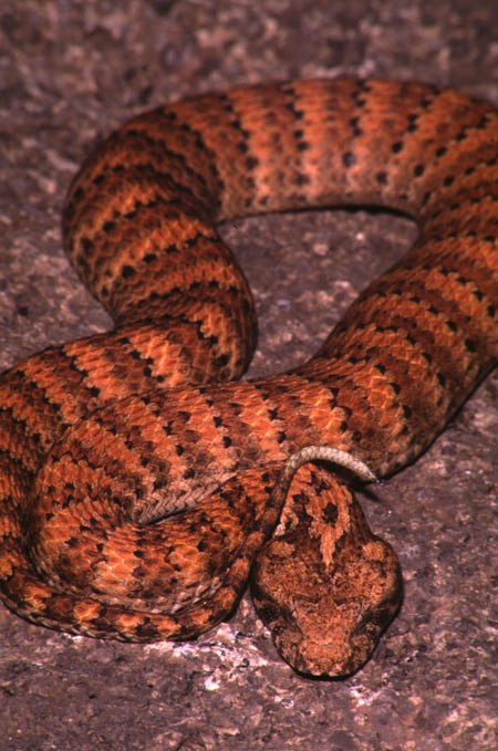
A SEVERE CASE OF STOMATITIS (CANKER OR MOUTHROT) IN A DEATH ADDER (ACANTHOPHIS ANTARCTICUS) ASSOCIATED WITH A REOVIRUS INFECTION
Raymond Hoser
488 Park Road
Park Orchards, Victoria, 3114, Australia.
Originally Published (with photo of affected snake) in Boydii, Autumn 2004:16-17.
ABSTRACT
Reported here is a case of Mouthrot in a snake believed to have been infected at the time with reovirus. The infection apparently appeared spontaneously (see the main paper for an explanation) and after a single cleaning and treatment healed completely. Whilst the exact cause of infection could not be definitively determined, it appears to have arisen at least in part due to underlying viral infection in the snake.
GENERAL
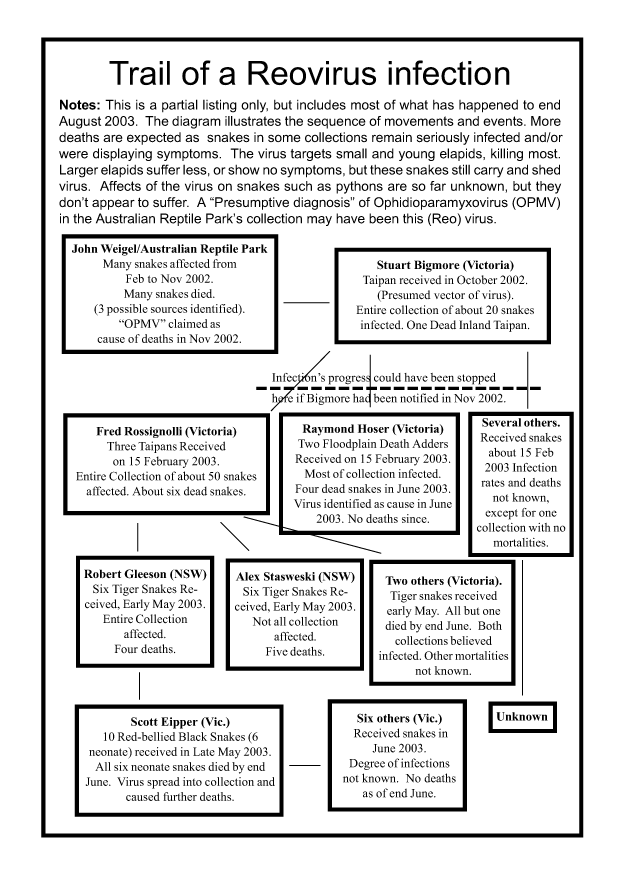
In June 2003, a die-off of four young neonate-sized death adders (Acanthophis spp.) in a group of eight occurred (Hoser 2003). All snakes were four months or less in age and between 20 and 28 cm in length. Three of the other death adders got ill and recovered.
The illness manifested with the following symptoms in the following order:
These signs led to a preliminary diagnosis of Ophidian Paramyxovirus, as the symptoms of this virus (in the literature) appeared to be identical (see Hoser 2003, Mader 1996 and references cited in both).
A account of this infection, including symptoms, diagnosis and other matters of relevance, including other affected collections, means and the pathways of infection was presented in Hoser (2003), the detail of which is not repeated here.
A longer paper detailing other aspects of the infection is anticipated.
Tests via electron microscopy at the Victorian Institute for Animal Science (VIAS) and the Commonwealth Scientific and Industrial Research Organisation (CSIRO) in Victoria by three scientists named at the foot of this paper, revealed the causative agent to be a previously unknown Reovirus.
Reoviruses are any of a group of RNA viruses including the rotavirus. Reoviruses do not harm all types of organism they infect. As a trend, those that do cause harm often cause greatest damage in young animals.
Reovirus has been known to cause death in venomous snakes outside Australia (see Mader 1996).
Other than a large Death Adder brought into the collection and later identified as the original carrier of the virus, five other large (adult sized) Death Adders were also exposed to the virus (via shared feeding tongs) and in the period from June 2003 to August the same year all were observed showing signs of infection.
The symptoms in these snakes were less acute than in the younger snakes and by the time all the snakes recovered in late August 2003, none had died.
In these snakes, symptoms manifested in the form of respiratory complaints, shown by either snout rubbing, visible exudate from the mouth or unnatural rubbing of exudate from the lower jaw as well as loss of appetite.
With the exception of two snakes (one of either sex) that would also open their mouth when breathing, the signs of infection were subtle and could easily have been missed if not specifically looked for.
This is because all snakes were large and over-fed, hence appetite loss on it's own wouldn't ordinarily have caused concern and the signs of respiratory ailment were also minimal and could have been easily overlooked.
The evidence of rubbing was only due to the clay-based substrate in the cage and had the substrate been paper, this evidence of rubbing would not have shown.
Evidence of rubbing showed up as encrusted dust around the mouth and snout regions and was also prominent in the affected neonate Death Adders.
On 1 July 2003 one of the adult-sized Death Adders (Acanthophis antarcticus) (then 65 cm total length) was fed a 3/4 sized dead rat (equal to about 7 large adult mice).
I then went away for 18 days before returning home on 19 July 2003 to inspect the snake.
To my surprise it had developed a large festering wound on the front lower jaw, slightly towards the left side. This was an archetypal mouthrot infection.
These are almost unknown in elapid snakes and hence my reporting of the case here.
In this case it was known with certainty that the wound was not caused by any injury from attack or feeding. This was because the snake had never eaten live food in its life and hence had never been attacked.
Furthermore a feeding Death Adder that experiences pain or resistance from sharp objects, such as leaves or twigs accidentally ingested will either move it's mouth around the object or if unable to remove the obvious pain, will regurgitate the food item with the offending object. Neither were observed at the time the snake ate the rat.
Noting that the snake had last been seen shortly after feeding on the rat and had externally presented as being in perfect condition, it was a rude shock to me to see this serious infection in the snake that had been left alone and untouched for nearly three weeks.
The infected snake was left untouched for another 24 hours, but the infection appeared to worsen quite substantially. By this stage the wound appeared to be weeping to an extent and collecting more dirt and other debris on it.
However from this point on the wound appeared to stabilize without treatment.
Notwithstanding this apparent stabilization, on 26 July 2003 I decided to clean the wound with warm water on a cotton wool bud, with several being used to clean the wound as best I could.
The wound was then treated with undiluted Betadine (Povidine Iodine) solution which was doused over the affected areas.
At the time of removing the crusted material from the front of the mouth and adjacent areas inside, the centre of infection was found to be a small crater like wound inside the jawline (as in behind the jaw, not facing out). Debris was also removed from this wound site leaving a small 'hole'. There was no evidence of the wound being created by any object, but that's not to say that one may not have been present at some time past.
It was before this clean-up of the infected wound that I photographed the infection by photographing the snake's head. The photograph only showed crusting around the edge of the jaw, as visible from an outside view and not the worst part of the infection and deep wound inside the jawline.
Noting that the snake was heading towards a sloughing cycle and the fact that handling of the snake tended to remove or damage outer scale layers, it was decided to leave the snake alone until after the slough unless the infection appeared to worsen.
Immediately after the slough which occurred on 30 July 2003, the wound was re-inspected and found to have improved markedly.
Notable here is that the snake sloughed in three pieces (not the usual one) and that some scales were retained on the neck. This result was undoubtedly a result of damage to scales caused at the time the snake had been held by the neck and handled on 26 July 2003.
More significantly however was that the snake did not defecate at any time during the sloughing process, including at least +/- 48 hours of the slough.
Non-defecation at time of sloughing in Death Adders is so rare as to be noteworthy, although on it�s own would not be regarded as cause for concern or alarm.
In this case it may have been indicative of underlying problems caused by the viral infection.
The improvement to the snake's mouth and general health obviously continued with the snake being observed sitting in a "normal" position and twitching it's caudal lure for the first time on 17 August 2003. Neither "normal" resting position or caudal luring had been observed prior to this date.
The snake was offered a small dead mouse on this date and she voluntarily took and ate it.
She took her first 3/4 sized rat on 21 August 2003, indicating a full recovery.
While there are a number of possible causes for the mouth infection as detailed here, it appears to be that at least in part, the Reovirus had a role.
The only likely explanation or reason as to how or why material could penetrate the inner lining of the jaw, is that such penetration could occur as the snake rubbed sticky exudate from it's mouth.
This rubbing is not natural behavior for Death Adders and in the captive situation has only been seen in terms of respiratory complaint associated with this Reovirus infection.
Under normal situations minor injuries or wounds do not become seriously infected festering wounds.
However it's known that one of the effects of viral infection is a decreased immune system making the affected animal more susceptible to secondary bacterial infections. This would include respiratory tract and mouth infections, including Canker as seen.
Canker associated with viral infections has been documented before.
However I think this case is the first such infection documented in an Australian elapid.
ACKNOWLEDGEMENTS
The female Death Adder referred to in this paper was bred by Alex Stasweski of Blacktown, NSW on 4 February 2002. It was held pursuant to relevant permits issued by the NSW and Victorian governments. The diagnosis of Reovirus in an affected Death Adder from the same collection was made by Mark Williamson of VIAS as well as Gary Crameri and Debra Middleton of the CSIRO.
REFERENCES
Hoser, R. T. 2003. OPMV in Australian Reptile Collections. Macarthur Herpetological Society Newsletter, Issue 38:2-8.
Mader, D. R. 1996. Reptile Medicine and Surgery. Saunders, Philadelphia, USA.
|
Herpetology papers index. |
Non-urgent email inquiries via the Snakebusters bookings page at:
http://www.snakebusters.com.au/sbsboo1.htm
Urgent inquiries phone:
Melbourne, Victoria, Australia:
(03) 9812 3322 or 0412 777 211